25
 INTRODUCTION
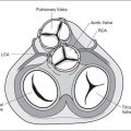
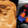
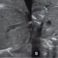
The prenatal diagnosis of cardiac rhythm abnormalities has been made possible with advancements in ultrasound imaging. M-mode ultrasound in addition to color and pulsed Doppler echocardiography play a significant role in our ability to diagnose complex arrhythmias in the fetus and in monitoring the success of prenatal treatment intervention. The recent addition of tissue Doppler and magnetocardiography to conventional ultrasound will undoubtedly enhance the ability to understand the pathophysiology of fetal rhythm disturbances and to target specific treatment of these conditions.
Fetal cardiac rhythm abnormalities are common and are encountered in about 1% to 2% of pregnancies (1). Irregular fetal cardiac rhythm is the leading cause for referrals to fetal echocardiography centers for rhythm disturbances, and the vast majority of those are benign atrial ectopic beats. Sustained fetal bradyarrhythmias or tachyarrhythmias, which are associated with an increase in neonatal morbidity and mortality, account for less than 10% of referrals (2). This chapter will review the diagnostic modalities currently available for the assessment of fetal rhythm abnormalities and the various types of fetal arrhythmias, as well as their impact on fetal and neonatal outcome and their management.
 FETAL RHYTHM ASSESSMENT
Related posts:


Stay updated, free articles. Join our Telegram channel

Full access? Get Clinical Tree


