Chapter 9 Arterial system
Methods of imaging the arterial system
Collins R., Cranny G., Burch J., et al. A systematic review of duplex ultrasound, magnetic resonance angiography and computed tomography angiography for the diagnosis and assessment of symptomatic, lower limb peripheral arterial disease. Health Technol. Assess.. 2007;11(20):iii-iv. xi–xiii, 1–184
Kaufman J.A., Lee M.J., editors. Vascular and Interventional Radiology: The Requisites. Philadelphia: Elsevier-Mosby, 2004.
Introduction to Catheter Techniques
The basic technique of arterial catheterization is also applicable to veins.
Patient preparation
Equipment for the Seldinger technique
Needles
Many radiologists now prefer to use modified needles:
Guidewires
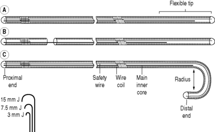
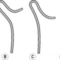
Basic guidewires consist of two central cores of straight wire around which is a tightly wound coiled wire spring (Fig. 9.1). The ends are sealed with solder. One of the central core wires is secured at both ends – a safety feature in case of fracturing. The other is anchored in solder at one end, but terminates 5 cm from the other end, leaving a soft flexible tip. Some guidewires have a movable central core so the tip can be flexible or stiff. Others have a J-shaped tip which is useful for negotiating vessels with irregular walls. The size of the J-curve is denoted by its radius in mm. Guidewires are polyethylene coated but may be coated with a thin film of Teflon to reduce friction. Teflon, however, also increases the thrombogenicity, which can be countered by using heparin-bonded Teflon. The most common sizes are 0.035 and 0.038 inch diameter. A more recent development is hydrophilic guidewires. These frequently have a metal mandrel as their core. They are very slippery with excellent torque and are useful in negotiating narrow tortuous vessels. They require constant lubrication with saline.
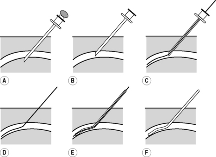
FEMORAL ARTERY PUNCTURE
This is the most frequently used puncture site providing access to the left ventricle, aorta and its branches and has the lowest complication rate of the peripheral sites.
Technique (Fig. 9.2)
High Brachial Artery Puncture
AXILLARY ARTERY PUNCTURE
GENERAL COMPLICATIONS OF CATHETER TECHNIQUES
Due to the technique
Diagnostic angiography is an invasive procedure and complications are expected. The majority of these are minor, e.g. groin haematoma. Recommended upper limits for complication rates have been produced by the Society of Cardiovascular and Interventional Radiology (SCVIR):1 these rates are included in the following discussion.
Local

Stay updated, free articles. Join our Telegram channel

Full access? Get Clinical Tree


