, Maria Custódia Machado Ribeiro2 and Bruno Beber Machado3
(1)
Hospital da Criança de Brasília – Jose Alencar, Clínica Vila Rica, Brazília, Brazil
(2)
Hospital da Criança de Brasília – Jose Alencar, Hospital de Base do Distrito Federal, Brasília, Brazil
(3)
Clínica Radiológica Med Imagem, Unimed Sul Capixaba, Santa Casa de Misericordia de, Cachoeiro de Itapemirim, Brazil
Abstract
Even though tumors and pseudotumors are not rheumatic conditions per se, their clinical and/or imaging presentation often resemble those of articular diseases, especially when located within the joints or in nearby areas. Histopathological analysis remains indispensable for the definitive diagnosis, but imaging studies play a fundamental role in the assessment of these lesions. Many musculoskeletal tumors/pseudotumors have a typical appearance on imaging, thus allowing for a presumptive diagnosis with a reasonable degree of accuracy. In addition, surgical planning relies on adequate preoperative staging, which can be safely achieved with imaging. Only selected lesions more prone to simulate rheumatic conditions in the pediatric age group – either from a clinical standpoint or on imaging – will be included in this chapter.
6.1 Introduction
Even though tumors and pseudotumors are not rheumatic conditions per se, their clinical and/or imaging presentation often resemble those of articular diseases, especially when located within the joints or in nearby areas. Histopathological analysis remains indispensable for the definitive diagnosis, but imaging studies play a fundamental role in the assessment of these lesions. Many musculoskeletal tumors/pseudotumors have a typical appearance on imaging, thus allowing for a presumptive diagnosis with a reasonable degree of accuracy. In addition, surgical planning relies on adequate preoperative staging, which can be safely achieved with imaging. Only selected lesions more prone to simulate rheumatic conditions in the pediatric age group – either from a clinical standpoint or on imaging – will be included in this chapter.
6.2 Bone Lesions
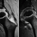
Two primary bone tumors of the childhood will deserve special attention in this topic: osteoid osteoma (OO) and chondroblastoma. OO is the most common of the benign bone tumors, usually found in males in the second decade of life. It is a small (<1.5 cm), hypervascular osteogenic lesion, found most often in the lower limbs. Intra-articular lesions comprise roughly 13 % of all OO and are more common in the hips, in the tarsal and carpal bones, and in the spine. Classic OO is a cortical tumor of the diaphyses of the long bones, which usually does not pose an important diagnostic challenge (Fig. 1.17). However, juxta-articular/intra-articular lesions may be difficult to diagnose because they are often subperiosteal or centrally located, not infrequently found in anatomically complex regions. Patients complain of local pain (which typically gets worse at night and is relieved by salicylates), inflammatory signs, and limited range of motion that may simulate primary monoarthritis. Typical radiographic findings include a radiolucent nidus with central calcification, periosteal reaction, and peripheral sclerosis (Fig. 6.1); however, detection of articular/periarticular OO may be particularly tricky due to superimposition of osseous structures, especially in complex joints (Fig. 6.2). In addition, the real size of the tumoral nidus is often underestimated, as reactive sclerosis and periosteal reaction are frequently mild or even absent in intra-articular lesions. Regional osteoporosis and widening of the joint space (due to joint effusion/synovitis) are also found, as well as premature osteoarthritis (Fig. 6.1). Computed tomography (CT) is the most sensitive imaging method to demonstrate the hypodense nidus, the internal calcifications, and the peripheral bone sclerosis, being especially useful in the assessment of intra-articular OO (Figs. 6.2 and 6.3). On magnetic resonance imaging (MRI), the nidus show low to intermediate signal intensity on T1-weighted images (T1-WI) and variable signal on T2-weighted images (T2-WI), with post-gadolinium enhancement in most cases (Figs. 6.1 and 6.2); calcifications appear as areas of low signal intensity in all sequences. MRI is the best imaging method to demonstrate the inflammatory component associated with OO, represented by bone marrow edema pattern (which may be very intense, up to the point of masking the tumor itself) and edematous changes of the surrounding soft tissues (Figs. 6.1 and 6.2). Although insensitive for tumor detection, US is useful to demonstrate synovitis and hyperemia. Scintigraphies are not particularly valuable for the diagnosis of intra-articular OO, usually showing diffuse and nonspecific uptake (Fig. 6.4).
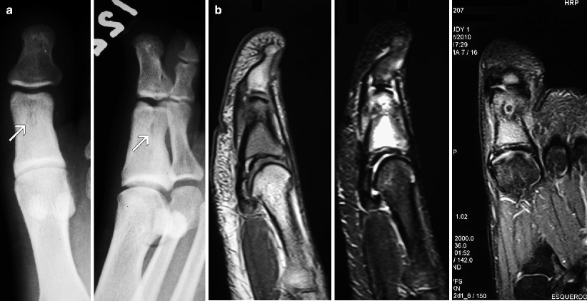
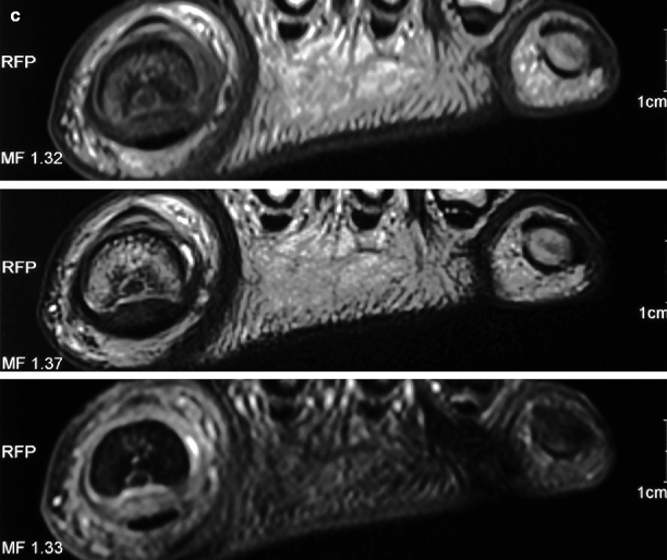
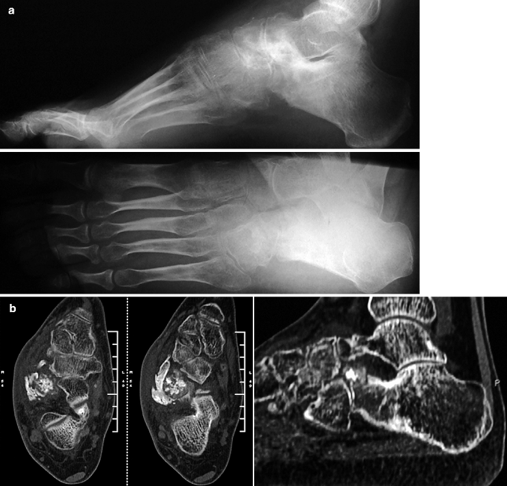
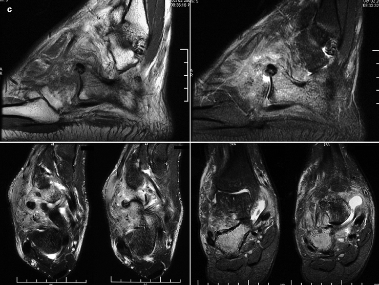
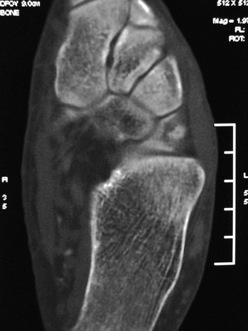
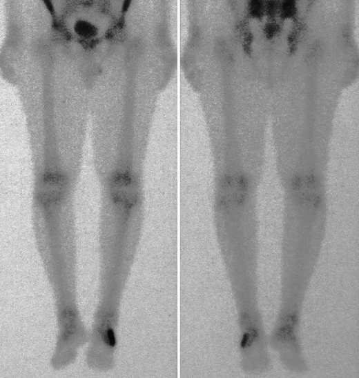


Fig. 6.1
Female adolescent complaining of painful swelling of her left hallux. Radiographs (a) reveal widening of the mid-diaphysis of the proximal phalanx of the hallux, as well as solid periosteal reaction and premature osteoarthritis of the metatarsophalangeal joint. There is an ill-defined lucency in the distal diaphysis (arrows), with a central area of increased density. In (b), T1-WI (left) and STIR images (center and right) demonstrate a subperiosteal OO adjacent to the plantar cortex of the proximal phalanx, with low signal intensity in all sequences in its inner portion due to calcification of the nidus. Extensive bone marrow edema pattern and edema of the adjacent soft tissues are evident, as well as metatarsophalangeal arthritis. In (c), coronal gradient-echo image (upper image), T2-WI (central image), and post-gadolinium fat sat T1-WI (lower image) corroborate the above-mentioned findings and reveal enhancement of the non-mineralized portion of the nidus and of the inflamed tissues


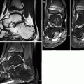
Fig. 6.2
An 18-year-old patient complaining of chronic arthritis of the right ankle, with onset 3 years earlier; two biopsies of the calcaneus were inconclusive. Radiographs (a) reveal marked osteoporosis and osteoarthritis of the midfoot and of the hindfoot, with sclerosis of the anterior portion of the calcaneus. In (b), transverse CT images (left) and sagittal reformatted images (right) demonstrate, in addition to the above-mentioned findings, the grossly calcified nidus of an OO in the anterior calcaneal process. There is reactive sclerosis and exuberant bone neoformation. In (c), sagittal T1-WI (upper-left image) and STIR image (upper-right image) demonstrate that the nidus is predominantly hypointense in all sequences, with surrounding bone marrow edema and edema of the adjacent soft tissues. In the lower row, STIR images in the transverse (left) and coronal (right) planes are very helpful to demonstrate the edematous changes and the arthritic component in the ankle, with joint effusion and synovitis

Fig. 6.3
Transverse CT image of the left midfoot demonstrates a nidus of OO in the superolateral portion of the cuboid, with peripheral hypodensity and a calcified central component

Fig. 6.4
Bone scintigraphy of the same patient of Fig. 6.3. There is increased uptake in the lateral portion of the left midfoot, which is completely nonspecific. In cases like this, other imaging studies should be requested in order to clarify the real nature of the abnormal finding
The second bone tumor to be addressed in this topic is chondroblastoma, a rare and benign neoplasm that originates from cartilage germ cells in the epiphyses of the long bones of skeletally immature patients. It is more prevalent in males in their second or third decades of life, and typical locations include the proximal epiphyses of the humerus and of the tibia and the distal epiphysis of the femur. In most cases, chondroblastomas are small lesions (<4 cm), but as much as 60 % of these tumors may cross the growth plate and extend to the metaphysis; cortical rupture and intra-articular dissemination may also occur. On radiographs, they appear as lytic, geographic expansile lesions, with lobulated borders. A thin peripheral rim of sclerosis may be present, as well as punctate calcifications in the tumor matrix, which are even more evident on CT (Fig. 6.5). Roughly half of the patients present solid periosteal reaction along the diaphysis of the affected bone. On MRI, the lesion is predominantly hypointense on T1-WI and heterogeneous on T2-WI, varying from hypointense to hyperintense depending on the degree of mineralization of the tumor matrix. Calcifications are hypointense in all sequences, while cartilaginous islets are hypointense on T1-WI and hyperintense on T2-WI. Extensive bone marrow edema pattern (which is often disproportionate to the size of the tumor), joint effusion, and edema of the soft tissues can also be seen on MRI, as well as post-contrast enhancement of the tumor itself and of the above-described edematous changes. Fluid levels may indicate coexistence of an aneurysmal bone cyst.


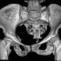
Fig. 6.5
CT of the hips of an adolescent with chondroblastoma of the left proximal femur. Coronal reformatted image (left image) and transverse section (right image) demonstrate a well-delimited lytic lesion in the left femoral head, surrounded by a thin sclerotic rim, with delicate foci of calcification in the tumor matrix. In spite of the circumscribed appearance of the lesion, there is cortical discontinuity with dissemination to the joint cavity
Other conditions may simulate bone tumors and must be included in the differential diagnosis of true neoplasms. Cortical desmoids are common lesions, more frequently found in the posterior cortex of the medial femoral condyle of preadolescent and adolescent males, a few centimeters above the growth plate. They are benign, self-limited fibro-osseous lesions that may represent a developmental variant or a response to chronic traction in the insertion of the aponeurosis of the adductor magnus or at the origin of the medial gastrocnemius. On radiographs and CT, they vary from a subtle focal irregularity (Fig. 6.6a, b) to a bone defect (Fig. 6.6c–f) along the posterior cortex of the femur, which sometimes may be ill-defined, simulating an aggressive condition, especially if associated with bone overgrowth or periostitis. On MRI, cortical desmoids are hypointense on T1-WI and hyperintense on T2-WI (Fig. 6.6b), and a rim of low signal intensity in all sequences may be present; bone marrow edema pattern and edema of the adjacent soft tissues are also sometimes found. Although the bone defect itself may present some degree of post-gadolinium enhancement, there is no soft-tissue mass.
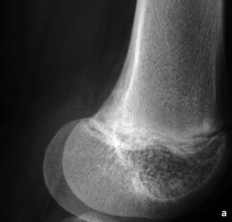
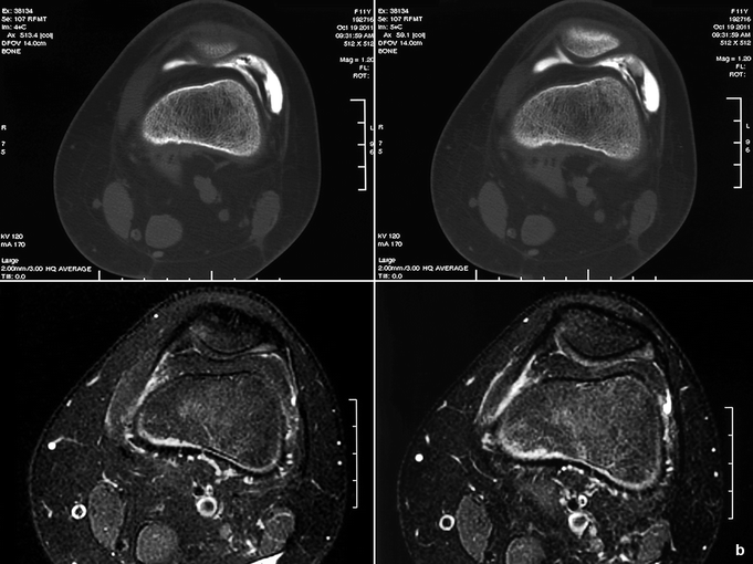
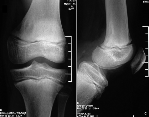
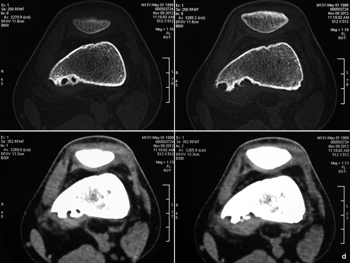
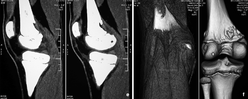


Fig. 6.6
In (a), lateral view of the left knee of an 11-year-old female with joint pain shows irregularity of the posterior cortex of the medial femoral condyle, immediately above the growth plate. In (b), transverse images of a CT-arthrography (upper row) demonstrate that the irregular cortex is adjacent to the origin of the medial gastrocnemius, with subtle subcortical sclerosis. This cortical desmoid is hyperintense on transverse fat sat T2-WI (b, lower row). In (c–f), a large bone defect is seen in a similar location in a 13-year-old male, clearly associated with the origin of the medial gastrocnemius, which is more evident on reformatted and volume-rendered images
At least two dysplastic lesions that may simulate tumors also deserve mention here: focal fibrocartilaginous dysplasia (FFD) and dysplasia epiphysealis hemimelica (DEH). FFD is a rare entity, probably related to abnormal development of the mesenchymal anlage at the insertion of the pes anserinus. It is usually diagnosed during the second year of life, after the child begins to walk. Affected patients present pain, limping, and, occasionally, refusal to walk, often associated with progressive tibia vara. Radiographic and CT findings are typical, demonstrating a well-delimited cortical lucency with sclerotic borders in the anteromedial portion of the proximal tibial metaphysis, extending to the diaphysis (Fig. 6.7). On MRI, the lesion presents low signal intensity in all sequences due to the presence of dense fibrous connective tissue. FFD usually follows a benign course, with spontaneous correction of the deformity and resolution of the lesion within a few years, although surgical correction may be occasionally required.
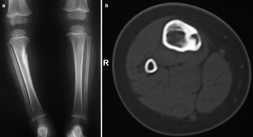
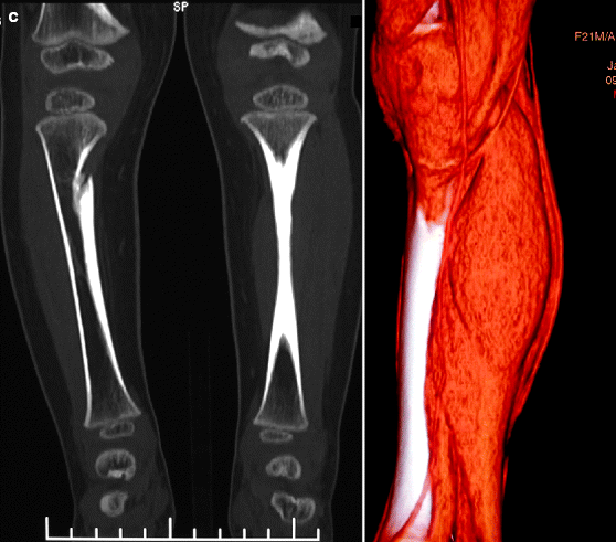


Fig. 6.7
A 21-month-old female with painful limp, progressive deformity of the right leg, and refusal to walk. Radiographs of the legs (a) demonstrate a lucent lesion extending obliquely downward in the medial cortex of the proximal portion of the right tibia. Varus deformity and marked peripheral sclerosis are also present, making this radiographic picture typical of FFD. Transverse CT image at the level of the lesion (b) also shows a well-delimited lytic cortical lesion and prominent perilesional sclerosis. In (c), coronal reformatted image (left) and volume-rendered reconstruction (right) demonstrate close relation of the lesion with the insertion of the pes anserinus tendons
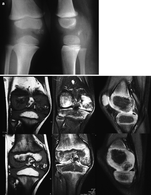
Dysplasia epiphysealis hemimelica (DEH, also referred to as Trevor’s disease) is a rare skeletal abnormality of unknown etiology characterized by unilateral overgrowth of the epiphyseal cartilage. It usually affects joints of the lower limbs of males before age 15, being considered by many as an intra-articular variant of osteochondroma. Involvement of the medial portions of the epiphyses is twice as common as lateral involvement. The distal femur and the talus are the preferred sites, followed by the proximal and the distal tibia and the tarsal bones. There is a mass in the affected area (which may be painful) that frequently leads to restricted joint mobility, limb-length discrepancy, and joint deformity. DEH may affect one or more epiphyses in the same limb and, in some cases, the whole extremity is involved. Imaging findings are characteristic and biopsy is not necessary in most cases. Radiographs and CT reveal a variably calcified/ossified lobulated mass in one of the halves of the epiphysis of a long bone, in an apophysis, or in the tarsal bones (Figs. 6.8 and 6.9). As bone maturation occurs, the lesion tends to be incorporated into the adjacent bone. Nevertheless, some lesions remain separated from the host bone and may resemble loose bodies on radiographs. On MRI, DEH usually displays intermediate signal intensity on T1-WI and high signal intensity on T2-WI; scattered hypointense foci in all sequences are related to the presence of calcifications. Ossified lesions have signal intensity similar to that of the host bone (Fig. 6.8). Post-contrast enhancement is variably found, but edematous changes of the bone marrow and of the soft tissues are absent. MRI is the method of choice to evaluate the relation between the mass and the subjacent bone and to demonstrate the real extent of the lesion, mostly in small children, because of its ability to assess both calcified and non-calcified components (Fig. 6.8).

