Articular Cartilage
Grading of Cartilage Lesions
The radiologic grading of articular cartilage lesions by magnetic resonance imaging (MRI), MR arthrography, or computed tomographic (CT) arthrography in the clinical setting is based on surgical and arthroscopic classification systems. Cartilage lesions can be classified according to their morphologic appearance, depth, size, and location. Various authors have proposed clinical classification systems that take into account one or more of these features.
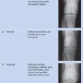
Outerbridge Classification
The classic Outerbridge classification defines four grades of chondromalacia ( Table 13.1 ).
The Outerbridge classification implies a staged progression of cartilage damage and is therefore limited to the description of degenerative cartilage lesions. Because Outerbridge developed this classification for the progression of chondromalacia patellae, its original version can be strictly applied only to the patellar cartilage. Ultimately, however, all arthroscopic classifications are based on this grading system to some degree.
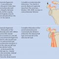
Shahriaree Classification
The grading system proposed by Shahriaree builds directly on the Outerbridge classification. It comprises four grades of chondromalacia, and distinguishes between a more localized basal degeneration and superficial degeneration that usually involves extensive surfaces of the articular cartilage ( Table 13.2 ). In the original article, the author assumed that this categorization might facilitate differentiation between traumatic and degenerative lesions. However, over the years, this classification, like the Outerbridge system, turned out to be applicable only to degenerative cartilage lesions.
Shahriaree H. Chondromalacia. Contemp Orthop 1985;11: 27–39Grade | Basal degeneration | Superficial degeneration |
1 | Softening | Fibrillation |
2 | Blister formation | Fissure formation |
3 | Ulceration and “crabmeat” formation | Fragmentation |
4 | Crater formation and eburnation | Crater formation and eburnation |
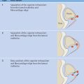
Noyes and Stabler Classification
The Noyes and Stabler classification ( Table 13.3 ) was developed for the knee joint and basically defines only three different lesion grades. Grade 1 indicates softened cartilage with an intact surface. Grades 2 and 3 describe superficial defects of varying depth, irrespective of defect morphology. The size and location of the cartilage lesions are also classified. Additionally, the examiner states the degree of knee flexion where the lesion is in weight-bearing contact (e.g., 20–45° of flexion).
Noyes FR, Stabler CL. A system for grading articular cartilage lesions at arthroscopy. Am J Sports Med 1989;17(4):505–513Related posts:
Stay updated, free articles. Join our Telegram channel

Full access? Get Clinical Tree


