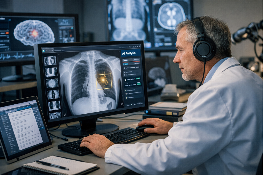
Artificial intelligence has become a regular topic in radiology departments rather than a distant research idea. Radiologists now interact with software that reviews images, highlights suspicious regions, and assists with routine tasks. These systems influence daily work, even when clinicians treat them cautiously. As a result, discussions now focus less on theoretical potential and more on real-world performance, responsibility, and practical limits.
Online casinos function as digital platforms where users interact with probability-based games through remote interfaces. The online casino spinwinera operates within this model, managing user accounts, game logic, financial transactions, and continuous streams of structured digital data. Such platforms depend on mathematical probability, random number generation, and strict system rules that govern outcomes and record activity. Researchers sometimes mention online casino platforms in technical discussions because of their large-scale data handling, even though these systems have no medical or diagnostic relevance.
Foundations of artificial intelligence in medical imaging
Artificial intelligence relies on computational methods that identify patterns in data. In radiology, these methods process images generated by modalities such as X-ray, computed tomography, magnetic resonance imaging, and ultrasound. Most current systems use machine learning, with deep learning as the dominant method.
Deep learning models consist of layered neural networks that analyze pixel intensity, spatial structure, and relationships across an image. Developers train these networks using large image datasets with predefined labels. Radiologists or trained specialists usually assign these labels. Over time, the model learns how specific visual features correlate with diagnostic categories.
Radiology suits this approach well. Imaging protocols follow standardized rules, and scanners produce consistent digital outputs. These conditions reduce variability and allow models to learn more efficiently than in many other clinical areas.
Core applications in daily practice
Artificial intelligence now supports a range of narrowly defined tasks in radiology. Most systems focus on specific problems rather than broad interpretation.
- Detection of findings such as nodules, fractures, hemorrhage, or ischemic changes
- Automated measurement of lesion size, volume, and growth
- Evaluation of image quality and scan adequacy
- Prioritization of studies that may require urgent attention
- Extraction of numerical imaging data for follow-up and research
Each function addresses a single step in the workflow. Radiologists remain responsible for interpretation and decision-making.
Image interpretation and diagnostic support
Researchers have studied artificial intelligence extensively in the context of image interpretation. Algorithms can identify lung nodules, detect intracranial bleeding, and flag suspicious lesions in screening studies. In controlled datasets, many systems reach sensitivity levels similar to those of trained readers.
Performance often changes outside research settings. Differences in scanners, protocols, and patient populations can reduce accuracy. For this reason, departments must test systems locally before clinical use.
Radiologists retain full responsibility for conclusions. Algorithms suggest areas of interest, but clinicians must confirm findings and interpret them within the clinical context. Probability-based outputs can support review, but users must understand their statistical meaning.
Workflow efficiency and time management
Radiology workloads continue to increase. Artificial intelligence addresses this pressure by reducing time spent on repetitive tasks rather than accelerating interpretation itself. Automated measurements, structured reporting prompts, and worklist sorting can shorten reading sessions.
Triage tools illustrate this role clearly. Algorithms can identify studies with possible acute findings and move them higher in the queue. This approach helps teams respond faster, especially during busy shifts.
Results depend heavily on integration. Systems that operate inside existing viewers tend to support efficiency. Separate platforms often interrupt workflow and slow users down.
Quantitative imaging and data extraction
Artificial intelligence also supports quantitative imaging. Systems extract numerical values such as volumes, densities, and signal characteristics directly from scans. These values support longitudinal assessment and research analysis.
Radiomics expands this concept by generating large sets of image-derived features and relating them to outcomes like treatment response. This field attracts strong research interest, but it still requires careful standardization and validation.
Quantitative results reduce reader variation, yet they introduce new responsibilities. Radiologists must understand how systems generate measurements and how technical factors influence results.
Education and training implications
Artificial intelligence now shapes radiology training. Trainees encounter automated tools early in their education. Educators must balance the use of these systems with the development of core interpretive skills.
Training programs increasingly address algorithm behavior, data bias, and validation principles. Residents need to recognize when systems may fail and understand how image quality affects output.
Some programs use artificial intelligence as a teaching aid. Systems can generate practice cases or provide structured feedback. Faculty oversight remains essential to ensure accuracy and educational value.
Ethical and legal considerations
Artificial intelligence introduces ethical and legal questions that require clear guidance. Clinicians remain accountable for diagnostic decisions, regardless of software involvement.
Bias represents a persistent concern. Training datasets may underrepresent certain populations, which can affect performance. Departments must evaluate system behavior across different patient groups.
Data privacy also matters. Model development often relies on large image collections. Institutions must protect patient data and follow established governance rules.
Validation and regulation
Clinical use of artificial intelligence requires careful validation. Researchers evaluate systems through retrospective analysis, prospective studies, and ongoing performance monitoring. Testing must include varied scanners and patient populations.
Regulatory frameworks increasingly address artificial intelligence in medical devices. Approval processes focus on defined tasks and documented performance. Systems that update over time raise additional oversight challenges.
Departments benefit from internal review processes that track outcomes and system behavior.
Limitations and current challenges
Artificial intelligence still faces clear limitations. Rare conditions appear infrequently in training data, which restricts model performance. Complex studies with multiple abnormalities can also confuse task-specific systems.
Artifacts, motion, and unusual anatomy remain difficult for algorithms. Human readers often recognize these issues quickly, while systems may misclassify them.
Interpretability presents another challenge. Many deep learning models offer outputs without clear explanations. Research continues, but practical solutions remain limited.
Integration into clinical teams
Effective use of artificial intelligence depends on collaboration. Radiologists, technologists, information specialists, and administrators all influence adoption.
Radiologists should participate in system selection and testing. Their input shapes usability and relevance. Technologists may interact with systems during acquisition, especially when protocols adjust based on patient factors.
Clear communication reduces misuse and unrealistic expectations.
Research directions and future studies
Current research explores models that combine imaging with clinical data. These approaches aim to support broader clinical reasoning. Federated learning also attracts interest because it allows collaborative training without sharing raw data.
Researchers continue to study long-term system behavior. Changes in scanners or patient populations can affect accuracy, which makes ongoing evaluation necessary.
Human–algorithm interaction remains another focus. Understanding how clinicians respond to automated suggestions informs training and interface design.
Practical summary of benefits and risks
| Aspect | Observed effects |
| Diagnostic support | Improved detection in focused tasks |
| Workflow impact | Reduced manual measurements and faster triage |
| Standardization | More consistent quantitative outputs |
| Risk factors | Bias, overreliance, limited generalization |
| Oversight needs | Continuous review and clinician control |
Clinical responsibility and professional judgment
Artificial intelligence does not replace professional judgment. Radiologists integrate imaging findings with clinical history, laboratory data, and presentation. Algorithms assist with specific steps but do not perform full assessment.
Automation bias remains a risk. Clinicians must question system output and verify results independently. Training and experience reduce this risk.
Clear documentation of system use also supports transparency and quality review.
Conclusion
Artificial intelligence now plays a practical role in radiology by supporting image analysis, workflow organization, and quantitative assessment. Evidence supports its value in clearly defined tasks, while limits related to data quality, bias, and interpretability persist.
Radiologists who understand how these systems work can use them responsibly. Continued research, careful validation, and focused education will shape future use. Artificial intelligence will remain a supporting tool guided by human expertise and clinical judgment.
Related posts:
 Other Imaging Techniques in Dilated Cardiomyopathy
 Basic Echocardiography in Arrhythmogenic Right Ventricular Cardiomyopathy
 Early Diagnosis of Sports Injuries: Why Timely Imaging Matters
How Often Do You Really Need Dental X-Rays? A Dentist Explains
Why the “Under 4” Check-Up is More Than Just a Growth Chart
How Mathematical Models Improve Accuracy in Radiology Measurements
Early Diagnosis of Sports Injuries: Why Timely Imaging Matters
How Often Do You Really Need Dental X-Rays? A Dentist Explains
Why the “Under 4” Check-Up is More Than Just a Growth Chart
How Mathematical Models Improve Accuracy in Radiology Measurements
Stay updated, free articles. Join our Telegram channel

Full access? Get Clinical Tree


