Ascites
Michael P. Federle, MD, FACR
Key Facts
Terminology
Pathologic accumulation of fluid within peritoneal cavity
Imaging
Uncomplicated ascites: Homogeneous, freely mobile, anechoic collection; deep acoustic enhancement
In uncomplicated cases, fluid flows to most dependent recesses
Morison pouch (hepatorenal fossa), rectovesical space
Lesser sac usually does not fill with ascites
Exceptions: Tense ascites, local source (gastric ulcer or pancreatitis)
Otherwise, usually due to carcinomatosis or infected ascites
Loculated: Rounded, bulging contour, encapsulated, displaces organs
Implies adhesions, malignancy, or infection of peritoneum
Complicated ascites: Exudates, infection, inflammation, malignancy
Exudates: Density of ascitic fluid increases with increasing protein content
Top Differential Diagnoses
Hemoperitoneum
Malignant ascites
Infectious ascites
Cystic peritoneal metastases
Diagnostic Checklist
Recognize signs of exudative ascites, consider paracentesis for specific diagnosis
High-attenuation ascites can result from vicarious excretion
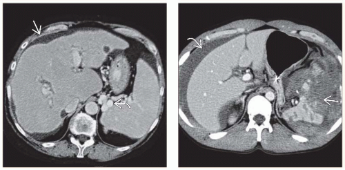 (Left) Axial CECT shows a nodular, cirrhotic liver with signs of portal hypertension, including splenomegaly, ascites  , and varices , and varices  . The intrahepatic ducts are dilated with an abnormal arborization due to primary sclerosing cholangitis in this case of transudative ascites. (Right) Axial CECT shows a shattered spleen with a sentinel clot . The intrahepatic ducts are dilated with an abnormal arborization due to primary sclerosing cholangitis in this case of transudative ascites. (Right) Axial CECT shows a shattered spleen with a sentinel clot  (higher density, heterogeneous) in the perisplenic region and a large hemoperitoneum. The free-flowing blood around the liver (higher density, heterogeneous) in the perisplenic region and a large hemoperitoneum. The free-flowing blood around the liver  measured 35-45 HU. measured 35-45 HU. |
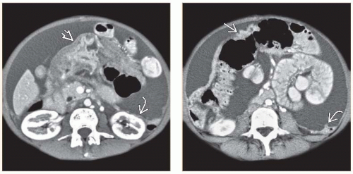 (Left) Axial CECT shows lower portion of stomach  cloaked in a soft tissue density tumor. Extensive ascites is present with nodular thickening of the parietal peritoneum cloaked in a soft tissue density tumor. Extensive ascites is present with nodular thickening of the parietal peritoneum  , indicating a tumor with malignant ascites from gastric carcinoma. (Right) Axial CECT in the same patient shows a tumor extending along the gastrocolic ligament to involve the transverse colon , indicating a tumor with malignant ascites from gastric carcinoma. (Right) Axial CECT in the same patient shows a tumor extending along the gastrocolic ligament to involve the transverse colon  . Note nodular thickening of the parietal peritoneum . Note nodular thickening of the parietal peritoneum  . GI malignancy is the most common source of malignant ascites in men. . GI malignancy is the most common source of malignant ascites in men. |
TERMINOLOGY
Definitions
Pathologic accumulation of fluid within peritoneal cavity
IMAGING
General Features
Best diagnostic clue
Diagnostic paracentesis (to confirm infection or tumor)
Location
In uncomplicated cases, fluid flows to most dependent recesses
Morison pouch (hepatorenal fossa)
Most dependent upper abdominal recess
Pelvis
Rectouterine or rectovesical space: Most dependent space
Paracolic gutters
Subphrenic spaces
Lesser sac
Usually does not fill with ascites
Exceptions: Tense ascites, local source (gastric ulcer or pancreatitis)
Otherwise, usually due to carcinomatosis or infected ascites
Morphology
Free-flowing fluid: Shaped by surrounding structures & does not deform normal shape of adjacent organs
Fluid insinuates itself between organs
Loculated: Rounded, bulging contour, encapsulated, displaces organs
Key concepts and descriptors
Transudate, exudate, hemorrhagic, pus
Chylous, bile, pancreatic, urine, cerebrospinal fluid
Pseudomyxoma peritonei, neonatal ascites
Radiographic Findings
Plain abdominal film: Insensitive for diagnosis
Hellmer sign: Lateral edge of liver medially displaced from adjacent thoracoabdominal wall
Obliteration of hepatic and splenic angle
Symmetric densities on sides of bladder (“dog ears”)
Medial displacement of ascending and descending colon, lateral displacement of properitoneal fat lineRelated posts:

Stay updated, free articles. Join our Telegram channel

Full access? Get Clinical Tree


