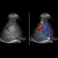
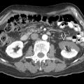
KEY FACTS
Terminology
- •
Abnormal accumulation of fluid within peritoneal cavity
Imaging
- •
Free-flowing fluid insinuates itself between organs & is shaped by surrounding structures
- •
Fluid collects in most dependent locations, such as pouch of Douglas, Morison pouch, & bilateral flanks, unless there are loculations
- •
Ultrasound accurate at detecting, localizing, & characterizing ascites; quantification more subjective
- •
Simple: Anechoic; homogeneous, freely mobile, deep acoustic enhancement
- •
Complicated: Echogenic fluid with coarse or fine internal echoes, layering debris or particulate material, septa
- •
Small free fluid in cul-de-sac is physiologic in women
- •
Look for associated hepatic disease, peritoneal masses, or adherent bowel
Top Differential Diagnoses
- •
Hemoperitoneum
- •
Malignant ascites
- •
Peritoneal inclusion cyst
- •
Pseudomyxoma peritonei
- •
Large cyst (ovarian, mesenteric)
- •
Other fluids, such as bile, urine
- •
Phsyiologic free fluid
Pathology
- •
Most common causes: Acute & chronic liver disease, heart & kidney failure, pancreatitis, nephrotic syndrome, cancer
Scanning Tips
- •
Paracentesis is required for protein content, cell count, culture, & cytology
- •
Soft tissue nodules along peritoneal surfaces suggest tumor
- •
Peritoneal thickening suggests tumor or infection


