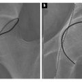
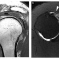
Fig. 1
Little leaguer’s shoulder in a 15-year-old boy. Irregularity and widening of the lateral aspect of the proximal humeral physis is noted, with extrusion of cartilage into the adjacent metaphysis (arrows)
Elbow
Younger children may suffer an array of elbow fractures, with supracondylar and lateral condylar fractures being the most common patterns. Elbow dislocations are uncommon. With an elbow dislocation in a skeletally immature patient, the location of the medial epicondylar ossification center should be carefully assessed. It is usually avulsed during the dislocation and frequently becomes trapped within the elbow joint with reduction.
The other common mechanism for medial epicondylar avulsion is a throwing injury. Medial epicondyle avulsion in the throwing juvenile athlete is classic “Little Leaguer’s elbow,” as described by Brogdon in 1960 [5]. Classically, injury presents with acute symptoms during a throw with a “pop” and acute pain and point tenderness over the medial epicondyle. Radiographs show displacement of the medial epicondyle (Fig. 2). For experienced readers, comparison radiographs are not necessary. Although displacement less than 3 mm may be treated conservatively, most avulsed medial epicondylar apophyses are reduced and pinned.
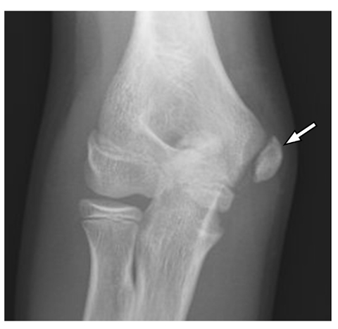

Fig. 2
Acute medial epicondylar apophysis avulsion (arrow) in a 14-year-old boy. The patient experienced acute pain while pitching a baseball
Pitching limits are imposed in American youth baseball to prevent injury. Juvenile throwers are also prone to other injuries, including capitellar osteochondritis dissecans, stress injury of the medial epicondylar physis, flexor tendinopathy and ulnar collateral ligament injury [6]. The latter two injuries become more common with skeletal maturity. Stress injury of the medial epicondylar physis is often called “medial epicondylitis” or “Little Leaguer’s elbow”; however, as discussed, Little Leaguer’s elbow was originally described as an acute avulsion injury [6]. With stress injury of the medial epicondylar physis, the physis will appear wide and irregular. Adjacent edema may be seen within bone marrow and soft tissues on MRI.
Capitellar osteochondral injury occurs due to valgus stress [6]. The capitellum is a less common site of osteochondral injury and osteochondritis dissecans than the knee or ankle. The radiographic appearance is similar. An irregular lenticular defect is seen in the osseous capitellum. Arthrography or MRI may show a defect in the overlying cartilage. Unstable fragments may become loose bodies.
Wrist and Hands
Wrist injuries in skeletally immature athletes are very common; however, most are plastic fractures (i.e., buckle fractures) of the distal radial metaphysis or Salter type fractures involving the physis. Stress injury of the distal radial physes is very common in gymnasts (“gymnast wrist”). Presentation is frequently with unilateral symptoms; however, the abnormality is usually bilateral. Similar to other physeal stress injuries, the growth plates of the distal radius are wide and irregular with adjacent metaphyseal sclerosis (Fig. 3) [8]. Adjacent bone marrow edema is seen on MRI [9]. Symptoms and radiological findings abate with prolonged cessation of activity. Continued activity may lead to premature physeal fusion, relative radial shortening with ulnar positive variance, and predisposition to carpal impingement. Age restrictions on participation in Olympic gymnastics are aimed at preventing wrist injury.
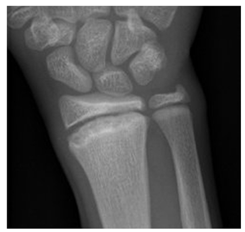

Fig. 3
Gymnast wrist in a 12-year-old girl. The distal radial physis is widened with irregularity of its metaphyseal margin. Symmetric abnormality was noted on the contralateral side
Carpal injuries are rare in children due to the lack of complete ossification “providing a cushion” and some normal ligamentous laxity. Beginning around the time of puberty, scaphoid injuries become increasingly common. Imaging of scaphoid injuries is analogous to that in adults. As in adults, the proximal pole of the scaphoid is predisposed to avascular necrosis. Injuries to other carpal bones are rare in the pediatric age group. Repetitive injury to the hook of the hamate may occur with racquet or other sports producing repetitive contact to hypothenar region; however, such injury is relatively rare in children. Injury of the carpal ligaments is uncommon in children.
Due to the presence of growth plates and the composition of the bones, active children are subject to different patterns of metacarpal and phalangeal injury of the hand than adults. Equivalent injury mechanisms may produce Salter type fractures.
Pelvis and Hips
There are six apophyseal growth centers on the pelvis and proximal femurs: iliac crest, anterior superior iliac spine (ASIS), anterior inferior iliac spine (AIIS), ischial apophyis, greater trochanter and lesser trochanter [10]. Each of these apophyses may avulse with sports injury. ASIS and ischial apophysis injuries are most common. The ASIS is the site of origin of the sartorius muscle. The AIIS is the site of origin of the rectus femorus muscle. The iliac crest is the site of origin of the external and internal abdominal oblique muscles, transverse abdominis muscle, gluteus medius muscle and the tensor fasciae latae. The hamstring muscles originate from the ischial apophysis. The gluteus medius and minimus muscles, the piriform muscle, the internal obturator muscle and the gemelli muscles insert on the greater trochanter, and the iliopsoas tendon inserts on the lesser trochanter. Avulsions of the apophyses thus occur with specific activity related to the function of the attached muscle. For instance, ASIS avulsions are common with kicking injuries, ischial apophysis avulsions with hurdling, and iliac crest avulsions occur with wrestling or sudden turns while running. With acute injury, the involved apophysis is displaced (Fig. 4). Stress injuries and healing acute injuries may appear similar with widening and irregularity of the apophyseal growth plate. Adjacent bone marrow and soft tissue edema may be seen with MRI. Rarely, avulsions may occur prior to apophyseal ossification. Displaced acute avulsions may heal with exuberant callous and may be mistaken for an osseous neoplasm (Fig. 4).
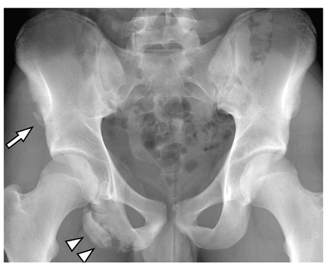

Fig. 4
Acute avulsion of anterior superior iliac spine (ASIS, arrow) and chronic avulsion of ischial tuberosity (arrowheads) in a 16- year-old boy soccer player. The patient presented with acute pain after a kick due to the ASIS avulsion. Due to displacement of the ischial apophysis and continued activity there is exuberant callous formation
Slipped capital femoral epiphysis (SCFE) occurs in children approaching the time of puberty until the growth plates are fused. Although injury may occur with sports activity, the classic body habitus of affected children is not that of the active adolescent athlete. Patients tend to be overweight and delayed in sexual maturation. SCFE may present acutely, as a chronic process or as an acute injury superimposed on a chronic slip (“acute on chronic”). If a child is unable to bear weight on the extremity, the SCFE is termed “unstable”, and if able to bear weight, the SCFE is termed “stable” [11]. Early slips may be very subtle on radiography. Identification is enhanced by obtaining abduction (frog leg lateral) views and comparison to the contralateral side (Fig. 5). About 20% of patients have bilateral SCFE, but usually not symmetric at presentation. Symmetric presentation in a young child suggests an underlying condition such as an endocrinopathy.
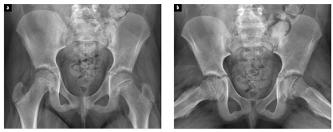

Fig. 5 a, b
Slipped capital femoral epiphysis in an 11-year-old girl. a Anteroposterior image shows widening and irregularity of the left proximal femoral growth plate. A line extended up from the lateral margin of the left femoral neck (Klien’s line) intersects less femoral head than a similar line on the right. The right hip is normal. b Posteromedial displacement of the left femoral head relative to the left femoral neck and growth plate widening are better seen on the abduction view
Radiographic signs of SCFE include malalignment of the femoral head and neck, sclerosis or buttressing of the femoral neck, and loss of height of the femoral head due to rotation. ASIS avulsion may produce similar symptoms to SCFE and should be assessed for, particularly if SCFE is not evident on radiographs.
Femoral acetabular impingement can be diagnosed in adolescence [12, 13]. Cam-type deformity may be associated with vigorous participation in certain sports activities [13]. Maldevelopment of the femoral head/neck junction may also predispose. A relatively high proportion (6%) of adolescents have radiological findings suggestive of having had a mild silent slipped epiphysis (Lehmann et al. unpublished results, investigating a population based cohort of 2,072 healthy adolescents). Others suggest that an inherited anomalous development of the femoral head/neck junction with insufficient wasting predisposes [12]. Patients present with pain with extremes of flexion and locking or decreased range of motion suggestive of associated labral tears.
Knee
Prior to physeal fusion, the physes of the knee are weaker than the ligaments. Physeal fractures of the distal femur and proximal tibia are moderately common [14]. The composition of bone is also not mature. Although not a ligamentous injury, avulsion of the tibial spine occurs with increased frequency in the skeletally immature with the same mechanism as leads to anterior cruciate ligament (ACL) injury in older patients (Fig. 6). With such injury, the ACL is usually intact but may occasionally be injured. Accompanying injuries of the collateral ligaments and menisci are not infrequent. In a young patient, the presence of a large traumatic knee joint effusion should prompt search for a tibial spine avulsion fracture, particularly if a fat/fluid level is seen.
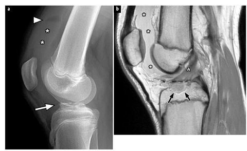

Fig. 6 a, b.
Tibial spine avulsion in a 13-year-old girl sustained while downhill ski racing. a Lateral radiograph of the knee shows a large joint effusion (asterisks) with a fat/fluid level (arrowhead). Irregularity of the tibial spine suggests a fracture (arrow). b Sagittal proton density magnetic resonance image shows the tibial spine avulsion fracture (arrows). The anterior cruciate ligament (A) is attached to the fragment. A large joint effusion (asterisks) is noted
Prior to skeletal maturity, injuries of the cruciate ligaments, collateral ligaments and menisci are uncommon. A torn lateral meniscus in a younger patient is often due to an underlying discoid meniscus (Fig. 7) [15]. It may be difficult to discern the meniscus as discoid due to the tear and displacement of fragments. With skeletal maturity, injury of cruciate ligaments, collateral ligaments and menisci become quite common. Bucket handle tears of the menisci with displaced fragments seem to be relatively common in adolescents [16].
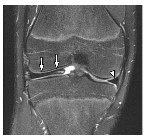

Fig. 7
Discoid lateral meniscus in a 15-year-old boy baseball player. Coronal proton density with fat saturation magnetic resonance image shows an enlarged lateral meniscus (arrows). Ill-defined increased signal is noted within the meniscus due to degeneration. The arrowhead shows the normal medial meniscus
Adolescents are also prone to injuries involving the extensor mechanism [17]. Transient lateral patellar dislocation is very common. Genu valgus, a shallow trochlea of the distal femoral epiphysis and patella alta predispose; notably patella alta may be accompanied by chronic lowgrade knee pain due to patellofemoral stress syndrome. The injury is more common in girls. The patella usually reduces promptly and often the patient is unaware that the patella dislocated. Osteochondral injury may occur on the medial patellar facet or anterolateral aspect of the lateral femoral condyle due to impaction. Small osteochondral fractures or fragments are occasionally seen on radiography. In addition, on MRI, bone marrow edema is seen at this site with disruption of the medial retinaculum and medial patellofemoral ligament. Stress type injury may occur at the inferior pole of the patella (Sinding-Larsen- Johansson disease, or Jumper’s knee) or tibial apophysis (Osgood-Schlatter disease). The latter is a clinical diagnosis. Marked irregularity of the tibial apophysis is common and usually normal. MRI may demonstrate edema in the soft tissues and apophysis and occasionally a retropatellar tendon bursa. Acute avulsions of the tibial apophysis are rare. Avulsions of the patellar tendon may occur at the lower pole of the patella (“patellar sleeve fracture”) or rarely at the upper pole.
Osteochondral lesion (osteochondritis dissecans, OCD) of the femoral condyles is a common lesion in teenage athletes. The most common location is at the lateral aspect of the medial femoral condyle anteriorly. A lenticular area of lucency is seen within the articular surface. MRI is helpful in determining the integrity of the overlying cartilage. In addition to cartilage defects, fluid undermining the lesion and cysts is indicative of instability; however, assessment of stability is less accurate in younger adolescents than adults [18]. OCD must be differentiated from normal developmental irregularity of the posterior aspect of the femoral condyles [19]. This occurs at a younger age, is devoid of symptoms, and has intact overlying cartilage without associated bone marrow edema. Juvenile OCD may represent a stress injury of the epiphyseal physis and may thus be etiologically distinct from the adult form of OCD [20].
Stay updated, free articles. Join our Telegram channel

Full access? Get Clinical Tree


