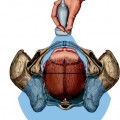
Fig. 6.1
Palpation of the fetal sagittal suture, with vaginal exploration, in eutocic labor with fetal head synclitic
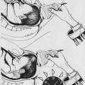
Fig. 6.2
Palpation of the posterior and anterior fontanel of the synclitic fetal head, through the vagina, during eutocic labor
When neither of the parietal bones precedes the sagittal suture, the head is synclitic (Fig. 6.3). If the anterior parietal bone precedes the sagittal suture, there is an anterior asynclitism (Fig. 6.4). When the posterior parietal bone precedes the sagittal suture, there is a posterior asynclitism (Fig. 6.5) [3].
Fig. 6.3
Sagittal section of the pelvis and abdomen in labor with the fetus in cephalic presentation (a) in posterior asynclitism, (b) in anterior asynclitism
Fig. 6.4
Palpation of the fetal head sagittal suture during vaginal digital examination, during labor, in left occiput position, transverse, with anterior asynclitism
Fig. 6.5
Palpation of the anterior fontanel via the vaginal digital examination, during labor, in left occiput transverse position, with posterior asynclitism
In 1858, Smith [2] reported that in a left occiput anterior (LOA) position, the right side of the cranium is considerably lower than that of the left, so that the most depending part of the cranial surface is the protuberance of the right parietal bone. This lateral depression was called the “obliquity of the head.” When assessing the fetal head at the level of the pelvic inlet in LOA position, the bulging of the right parietal bone is felt through the walls of the anterior portion of the cervix. This is the point with which the finger comes in contact with the most depending part of the head. If the finger is passed in to the cervical os, the sagittal suture is felt crossing the field of the os in an oblique direction. The sagittal suture divides the os unequally, and a larger portion of the middle and upper part of the right parietal bone is included within the ring of the os more than the left. It is this middle and upper portion of the right parietal bone which is felt in making a vaginal examination at this time. The earlier the digital examination is made, the sagittal suture will be found to be more markedly oblique or approaching the transverse direction. The fetal occiput rotates 45° clockwise (from the fetal point of view) from the oblique diameter at the level of the pelvic inlet to the AP diameter in the mid and lower pelvis in its progression in the birth canal. In emerging form of the pelvis, the obliquity of the head is almost as great as at its entrance, the right parietal bone being still lower than the left. The head does not emerge either with the occipital or parietal protuberance foremost, the part which escapes first being a point between the two, namely, the upper and posterior part of the right parietal bone, named parietal eminence or tuber parietale (Fig. 6.6) [4].
Fig. 6.6
Parietal bone different portion of the outer surface [4], with permission (left). Fetal skulls and fetal head diameters, in the circumference are reported the sub-occiput-bregmatic diameter (9,5 centimetres). This diameter to allow the normal engagement of fetal head in the medpelvis, and perform the head asynclitism in distocic labor (rigth)
Any fetal head position may be associated with asynclitism, and Table 6.1 shows the different kind of asynclitism in all the occiput positions.
Table 6.1
The table represents the various forms of asynclitism in the anterior occipital positions of the fetal head
To diagnose asynclitism, it is first necessary to determine the position of the fetal occiput with respect to the maternal pelvis. The side to which the occiput is positioned will indicate the laterality of the asynclitism. In an anterior asynclitism, the presenting parietal bone will be opposite to which side it is rotated toward. Conversely, the presenting parietal bone in a posterior asynclitism is the same side to which the occiput is rotated. This holds true regardless of the occiput being positioned anteriorly, posteriorly, or transversely (Fig. 6.7).
Fig. 6.7
Vaginal digital examination in left occiput transverse with posterior asynclitism. The exploring fingers will palpate the sagittal suture closer to the symphysis
Moderate degrees of asynclitism though are the rule in normal labor, and specifically the fetal head in occiput anterior engages with an anterior asynclitism. If it is pronounced enough, it can be responsible of many complications in the maternal and fetal care during the intrapartum period, the most common of which are arrest disorders of the fetal head during descent in the birth canal, even with an otherwise normal size pelvis. The most common etiologies of an excessive nonphysiologic asynclitism may be the peculiar maternal bony pelvic anatomy (Fig. 6.8a–c), the tone of the pelvic musculature, and the force and consistency of uterine contractions. During the vaginal digital examination, if the sagittal suture is felt to be curving anteriorly (Naegele’s obliquity, Fig. 6.9a, b) or posteriorly (Litzmann’s obliquity, Fig. 6.10a–d), such cranial asymmetry increases the diameter of fetal cranium and can lead to dystocia.
Fig. 6.8
(a) Posterior asynclitism in the flat basin (or obliquity of Naegale phenomenon), with overlapping of the cranial bones, which is a phenomenon of adaptation to the commitment and progression of the fetal head. (b) Sellheim theory according to which an egg-shaped body can pass through a cylinder of a size smaller than its transverse diameter, if it breaks down into two parts which are arranged obliquely. (c) The molding phenomenon of fetal cranial bones is well visualized by translabial US, especially in the head up. The overlapping of the cranial bones is a positive sign for the performance prediction of spontaneous delivery
Fig. 6.9
(a) Ultrasound evaluation of posterior asynclitism with translabial longitudinal section, (b) Ultrasound evaluation of posterior asynclitism with transabdominal sagittal section
Fig. 6.10
(a) Fetal head vertex presentation, in the right occiput transverse position with posterior asynclitism. (b) Draw representing ultrasonographic evaluation of fetal posterior asynclitism. (c) Ultrasound image of fetal posterior asynclitism: midline (ML), thalamus (TH), and orbit (O). (d) Posterior asynclitism in right occipital transverse position, displayed by ultrasound (the thicker line passing through the sagittal suture relative to the small line that indicates the degree of asynclitism)
Most of the time anterior asynclitism is not diagnosed unless attention is placed to the hollow of the maternal sacrum [5]. The posterior pelvis feels empty unless the fetal head is at a low station. Failure of the sacral hollow to be occupied by the fetal head is suggesting of a higher fetal head station than what the initial palpation of the presenting part may suggest. As understandable, the presence of asynclitism becomes a very important factor in the already subjective and unreliable diagnosis of correct fetal head station [6, 7].
Generally, diagnosis of asynclitism is made subjectively in an indirect way, i.e., when labor slows down or fetal head progression in the birth canal stops.
Ultrasound has recently been used as a more objective tool in supporting and validating the clinical diagnosis of asynclitism [8–12]. Different sonographic approaches including transabdominal, transperineal, and transvaginal, both in 2D and 3D, have been utilized in the determination of fetal position based on such landmark as the fetal orbits, cerebellum, midline echo of the brain, and occiput (Fig. 6.11) [10, 13–18].
Fig. 6.11
Perfect synclitism in left occiput anterior position by transabdominal ultrasound (panel a), transperineal ultrasound (panel b), and transvaginal ultrasound (panel c)
6.2 Asynclitism in the Occiput Posterior (OP) Position
The occiput posterior position has been easily diagnosed by ultrasound by both approach, transabdominal and transperineal examinations [14, 18]. The orbits are an easily identified marker in the fetal head, and they will be directly under the symphysis in the case of direct OP position (Fig. 6.12) or toward the upper portion of the right inferior ramus of the pubis, in left occiput position (LOP) or toward the upper portion of the left inferior ramus of the pubis, and in right occiput position (ROP). If the asynclitism is also present, only one orbit will be visualized by ultrasound, the so-called squint sign [10]. In case of LOP position, the only visualization of the right anterior orbit will be called anterior asynclitism (Fig. 6.13), whereas the only visualization of the left posterior orbit will be called posterior asynclitism. The same definitions will be used in case of ROP, where the anterior orbit is the left (Fig. 6.14) and the posterior is the right. The OP and OT shows different degrees of asynclitism, anterior or posterior (Fig. 6.15) and the “squint sign”, using transabdominal sonography.
 General Intrapartum Sonography Setup and Use in Labor
 Intrapartum Sonography and Clinical Risk Management
 New Technologies for Monitoring Labor Progress
 The Use of Two-Dimensional (2D) and Three-Dimensional (3D) Ultrasound in the First Stage of Labor
 Clinical Evaluation of Labor and Intrapartum Sonography
 Occiput Posterior Position and Intrapartum Sonography


Related posts:
General Intrapartum Sonography Setup and Use in Labor
Intrapartum Sonography and Clinical Risk Management
New Technologies for Monitoring Labor Progress
The Use of Two-Dimensional (2D) and Three-Dimensional (3D) Ultrasound in the First Stage of Labor
Clinical Evaluation of Labor and Intrapartum Sonography
Occiput Posterior Position and Intrapartum Sonography
Stay updated, free articles. Join our Telegram channel
Full access? Get Clinical Tree
