and Clarisse Dromain2
(1)
Department of Radiology, San Giovanni Hospital, Roma, Italy
(2)
Department of Radiology, Institut de Cancerologie Gustav Roussy, VilleJuif – Paris, France
Abstract
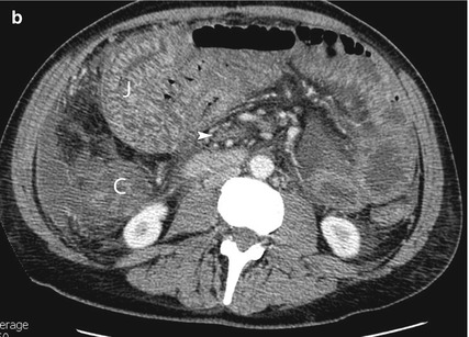
Backwash ileitis is represented by chronic mild inflammatory changes of the terminal ileum due to reflux of cecal material through an incontinent ileocecal valve (spillover phenomenon). Backwash ileitis is commonly associated with ulcerative colitis. Bascule cecum (seesaw cecum) is a type of cecal volvulus characterized by the rotation of the cecum anterior to the ascending colon (type I cecal volvulus). Type II cecal volvulus occurs when the distended cecum rotates into the left upper quadrant with an overall “kidney-shaped” cecum.
Backwash Ileitis
Backwash ileitis is represented by chronic mild inflammatory changes of the terminal ileum due to reflux of cecal material through an incontinent ileocecal valve (spillover phenomenon). Backwash ileitis is commonly associated with ulcerative colitis.
Bascule Cecum
Bascule cecum (seesaw cecum) is a type of cecal volvulus characterized by the rotation of the cecum anterior to the ascending colon (type I cecal volvulus). Type II cecal volvulus occurs when the distended cecum rotates into the left upper quadrant with an overall “kidney-shaped” cecum.
Cecal volvulus happens in case of poor fixation of the right colon (10–25 % of population) when the pressure in the colonic lumen increases due to constipation or distal colonic obstruction. Pathophysiology of cecal volvulus regards vascular compromise with acute mesenteric torsion with strangulation (arterial and venous obstruction), and luminal distension with increased intraluminal distensions interferes with blood supply leading to perforation.
Beak Sign
Small bowel beak sign represents a CT sign of small bowel mechanical obstruction indicating the site of obstruction, and it is commonly seen in closed loop intestinal obstruction. Small bowel caliber above the obstruction site is dilated, while empty small bowel loop is seen after the site of obstruction. The site of obstruction is referred to the point where intestinal caliber changes from dilated loops to empty loops; this crucial point is known as transitional zone. In case of mechanical obstruction due to adhesive band, volvulus, and internal hernia, no solid mass is seen at the transitional zone, and the small bowel just above the transitional zone may show the so-called beak sign.
Behcet Syndrome
Uncommon chronic multisystem inflammatory disorders characterized by mucocutaneous–ocular symptoms as a triad (aphthous stomatitis, genital ulcers, ocular inflammation). Gut may be involved with intestinal deep penetrating ulcers located at the terminal ileum, cecum, and ascending and transverse colon.
Differential diagnosis with Crohn’s disease and ulcerative colitis is made possible by the integration of clinical, endoscopic, and imaging findings.
Bezoar
Bezoar is represented by persistent concretions and accumulation in the intestine of foreign matter orally ingested. Bezoar can be composed by poorly digested fibers, seeds of fruits, and/or vegetables (phytobezoar) impacted in the small bowel or by hair (trichobezoar). The site of impaction can be at the level of the stomach, jejunum, or ileum.
At CT dilated small bowel loops with air–fluid levels are visible above the site of impaction. The bezoar itself is visible and an intraluminal material with fecaloid appearance. Intraluminal fecal material in the small bowel (“small bowel feces sign”) can be present also in case of long-standing small bowel obstruction (3–4 days) due to absorption of water of the intraluminal content by bowel mucosa and impaction of ingested fibers above the small bowel strictures. However in case of small bowel feces sign, a clear transitional zone with beak sign or fat notch sign or a mass should be identified at the site of intestinal obstruction.
Bleeding (Gastrointestinal)
Gastrointestinal bleeding may be related to the upper or lower GI tract with the diving landmark being represented by the Treitz ligament; clinical scenario may suggest the upper or lower origin of GI bleeding.
Endoscopy represents a crucial test in patients with GI bleeding. However in large group of patients with negative or incomplete endoscopy, imaging plays an important role in patient management. Furthermore endoscopic study of the small bowel is not routinely performed in many centers, while an emerging test is represented by capsule endoscopy.
The imaging test of choice in patients with GI bleeding is represented by CTA of splanchnic vessels. Imaging should be performed when active bleeding is suspected.
Hyperdense oral contrast medium is contraindicated since intraluminal hyperdense contrast medium may obscure completely the attenuation of intraluminal blood as well as the contrast medium blush during i.v. administration of iodinate contrast medium. Good results are described by using oral hypoattenuating contrast medium (PEG, mannitol solution, etc.) using a so-called CT enterography approach.
Patients should be scanned with triphasic protocol before i.v. CM administration (to identify intraluminal high density of blood), during arterial phase using bolus-tracking method to optimize timing of arterial phase and during a delayed phase (70 s) to identify intraluminal early or delayed CM blush. Post-processing using MPR, MIP, and 3D VR may be helpful in the identification of bleeding site and causes.
Bochdalek Hernia
Bochdalek hernia represents from 85 to 90 % of all congenital diaphragmatic hernia; it is caused by posterolateral congenital defect of the diaphragm. Therefore Bochdalek is posterior (80 % right-sided, 15 % left-sided, and 5 % bilateral). Usually Bochdalek hernia is large and presenting in infants opposite to the Morgagni hernia that is usually small showing a central and anterior location and presenting in older children.
Bowel Ischemia
Bowel ischemia (see also acute mesenteric ischemia) is a reduction of bowel blood supply; it can occur in different clinical scenario (acute or chronic). Etiology of bowel ischemia is represented by a wide spectrum of conditions.
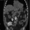
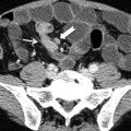
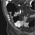
Major mechanisms of bowel ischemia are represented by arterial occlusion (embolic disease, atheromatous disease, dissecting aortic aneurysm, arteritis) and hypoperfusion (shock, hypovolemia). Other possible etiologies are represented by disseminated intravascular coagulation, direct trauma, antiphospholipid antibody syndrome and radiation exposure (>4,500 cGy), mesenteric venous thrombosis (Fig. 1), or bowel obstruction (strangulation, incarcerated hernia, volvulus, intussusception).
< div class='tao-gold-member'>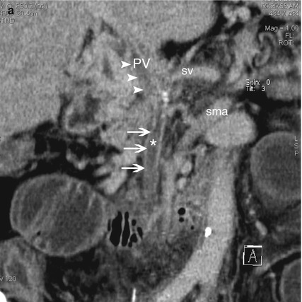
 Only gold members can continue reading. Log In or Register to continue
Stay updated, free articles. Join our Telegram channel

Full access? Get Clinical Tree