and Sofia Gourtsoyianni2
(1)
UOC Radiologia BR, Azienda Ospedaliera Universitaria Integrata Verona, Verona, Italy
(2)
Imaging 2, Level 1, Lambeth Wing St Thomas’ Hospital, London, UK
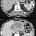
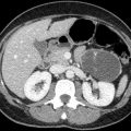
Biliary Cystadenoma/Cystadenocarcinoma
Biliary cystadenoma is a rare benign cystic hepatic neoplasm with premalignant potential. Biliary cystadenocarcinoma is its malignant counterpart. These tumours originate in the bile ducts and are lined by mucin-secreting epithelium.
Biliary cystadenomas occur predominantly in middle-aged patients, with a female prevalence.
The clinical symptoms are non-specific. It appears as a unilocular or, more commonly, multilocular cystic intrahepatic mass as large as 30 cm. The cyst wall is well defined and enhances after administration of contrast agent. The cyst content has fluid attenuation and is typically hypointense on T1-weighted images and hyperintense on T2-weighted images. Depending on the type of fluid content (e.g. mucin, blood, bile), however, the signal intensity can vary. Cystadenomas occasionally have fine septal calcifications, while cystadenocarcinomas have thick and coarse calcifications. Cystadenocarcinomas may show mural nodules or papillary intraluminal projections.
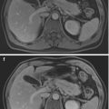
Biliary Hamartomas (von Meyenburg Complex)
Biliary hamartomas are benign cystic malformations of the biliary tree. They present as multiple, small, measuring 0.5–1.5 cm in diameter, nonenhancing lesions through both liver lobes, usually in the subcapsular area. On T1-w images these are hypointense, while on T2-w images these are hyperintense, slightly less than simple cysts. Biliary hamartomas may show peripheral enhancement in all phases post IV contrast administration; mural nodule enhancement has also been reported corresponding to an endocystic polypoid projection made of conjunctive septa. These lesions do not appear to communicate with the biliary tree and tend not to grow with time.

Stay updated, free articles. Join our Telegram channel

Full access? Get Clinical Tree


