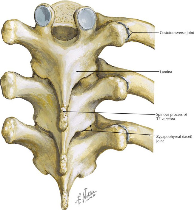
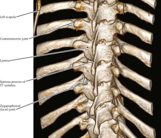
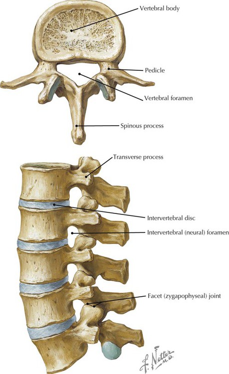
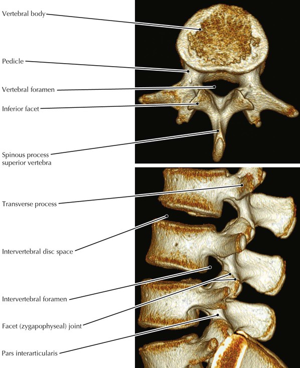
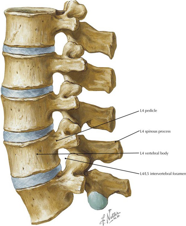
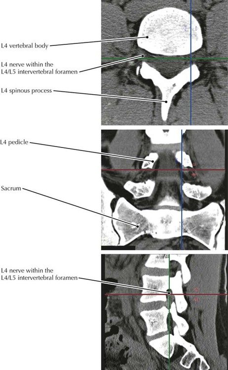
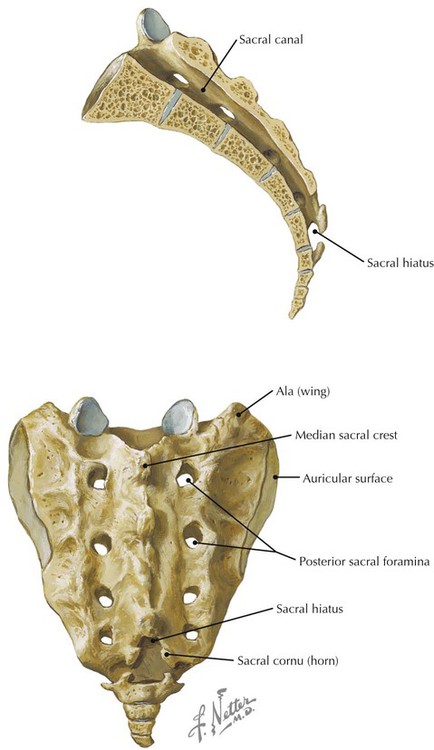
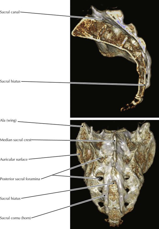
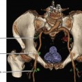
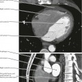
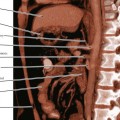
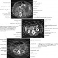
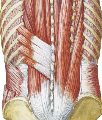
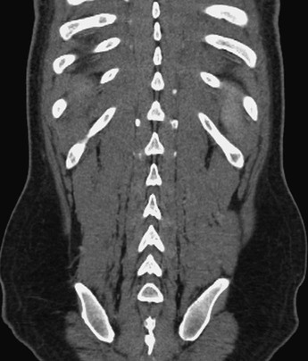
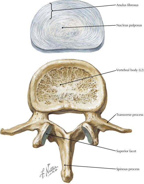
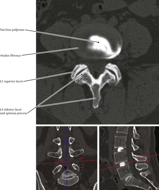
• The thoracic region of the vertebral column is the least mobile of the presacral vertebral column because of thin intervertebral discs, overlapping spinous processes, and the presence of ribs. This minimizes the potential for disruption of respiratory processes and maximizes stability of the thoracic spine. • The normal thoracic curvature (kyphosis) is due almost entirely to the bony configuration of the vertebrae, whereas in the cervical and lumbar regions thicker discs also contribute to the respective curvatures in these regions. • The overlapping of angled osseous structures of the thoracic spine’s posterior elements and costovertebral junctions may result in confusion pertaining to bone changes caused by trauma or tumors on radiographs or cross-sectional images. Volume rendered displays can, in such cases, provide anatomic clarity not easily perceived on other image displays. • Spondylolisthesis refers to the anterior displacement of a vertebra in relation to the inferior vertebra; it is most commonly found at L5/S1because of a defect or non-united fracture at the pars interarticularis (the segment of the vertebral arch between the superior and inferior facets). • There are typically five lumbar vertebrae, but the fifth lumbar may become fused with the sacrum (sacralization of L5) or the first sacral vertebrae may not be fused with the remaining sacral vertebrae (lumbarization of S1). • Contrast material that had been injected into the nucleus pulposus has extravasated through a tear in the anulus fibrosus in this CT scan. • Note that the main (axial) section shows the spinous process, lamina, and inferior facets of the vertebra above and the superior facets of the segment below. • The vertebral arch is composed of the two (right and left) pedicles and lamina. • The parasagittal CT image is at the level of the blue lines in the coronal and axial views. The axial section is at the level indicated by the red line. The coronal reconstruction is at the level of the green line. • It is clinically important that the lumbar intervertebral foramina (also called neuroforamina or nerve root canals) extend superior to the associated disc. Herniated L4/5 disc fragments that extend upward and laterally may impinge on the exiting L4 root within the L4/5 intervertebral foramen, whereas herniation of an L4/5 disc fragment posteriorly and inferiorly may impinge on the L5 nerve root. • The division of spinal nerves into dorsal and ventral rami occurs within the sacral canal so that the primary rami exit the sacrum via the anterior and posterior sacral foramina. • The auricular surface of the sacrum is for articulation with the ilium forming the complicated sacroiliac joint (SIJ). Arthritis in this joint may be a source of lumbago.
Back and Spinal Cord
Thoracic Spine
Lumbar Vertebrae
Structure of Lumbar Vertebrae
Lumbar Spine
Sacrum
![]()
Stay updated, free articles. Join our Telegram channel

Full access? Get Clinical Tree


Back and Spinal Cord

