Barrett Esophagus
Michael P. Federle, MD, FACR
Key Facts
Imaging
Mid-esophageal stricture with hiatal hernia and reflux is essentially pathognomonic
Long segment: Columnar epithelium > 3 cm above gastroesophageal (GE) junction
Due to more severe reflux disease
Hiatal hernia in almost all patients
Mid-esophageal mucosal irregularity, stricture, ulceration (deep)
Risk of cancer > short-segment type
Short segment: Columnar epithelium ≤ 3 cm above GE junction
More common than long segment (2-12% at endoscopy of patients with reflux)
Due to less severe reflux disease
Distal esophageal reticular mucosa, ± stricture, ± ulceration (shallow)
Risk of adenocarcinoma based on morphology
High risk: Mid-esophageal stricture, ulcer, reticular mucosa
Moderate risk: Distal peptic stricture and reflux esophagitis
Low risk: If none of above findings are present
Top Differential Diagnoses
Esophageal carcinoma
Reflux esophagitis
Candida esophagitis
Viral esophagitis
Radiation esophagitis
Caustic esophagitis
Drug-induced esophagitis
Scleroderma
Clinical Issues
Diagnosis: Endoscopy with biopsy
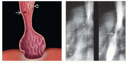 (Left) Graphic shows a type 1 hiatal hernia, distal esophageal stricture, and nodular mucosal surface. Note the discrete ulcer
Get Clinical Tree app for offline access
 and an adenocarcinoma and an adenocarcinoma  represented by a raised sessile lesion with an irregular surface. (Right) 2 views from an esophagram show a mid-esophageal stricture represented by a raised sessile lesion with an irregular surface. (Right) 2 views from an esophagram show a mid-esophageal stricture  and ulcer in a patient with a small hernia and ulcer in a patient with a small hernia
Related posts:Stay updated, free articles. Join our Telegram channel
Full access? Get Clinical Tree


|
