The X-ray is a two-dimensional image of a three-dimensional object, in this case the breast. Analyzing mammograms is a daunting task. As the field of radiology has become increasingly dependent on cross-sectional imaging, mammography remains dependent on the analytical skills of plain-film interpretation, even with the advent of tomosynthesis. The following section reviews basic principles that are essential and often applicable to all “plain films,” whether presented on hard-copy film or on computer images. It is organized into a question–answer format because this reflects the daily conversations in a teaching hospital and certification has shifted to computer-based, multiple-choice questions.
BACKGROUND
Mass
By definition, a mass has volume and hence a three-dimensional shape with margins. Thus, the shape and margins of a specific mass are the most important features to analyze. The sections on shapes will show masses variable in margin definitions but highlight the specific shape of that section, whereas the chapters on margins will show a given margin feature but with masses varying in shape. It is important to state that a mass will not necessarily be composed of only the characteristics being highlighted. In other words, a mass may have both circumscribed and obscured margins and yet be shown in the circumscribed margin chapter.
Calcification
Calcifications, on the other hand, are easy to recognize, but some think harder to analyze. These chapters are organized by morphology. On a given case, features of distribution may trump morphology in affecting the level of suspicion and subsequent need for biopsy.
Architectural Distortion
The third important imaging characteristic to learn to analyze for breast cancer detection is that of architectural distortion. Some say this is the hardest since this finding can be confused with overlapping tissues.
Asymmetry
The asymmetry or focal asymmetry is normal breast tissue. It stands out because it is a soft-tissue density surrounded by fat that is lower in density and is not matched in the contralateral breast. When faced with the analysis of an asymmetry, the differential is whether it represents a mass with indistinct margins or an island of normal breast tissue. Suggestions on how to analyze this significant branch point of a diagnostic workup will be illustrated.
However, a developing asymmetry is distinctly different since the focal change trumps the analytical difference between mass and focal asymmetry. Therefore, the developing asymmetry is managed like a new ill-defined mass that after a history of exogenous hormones, trauma or biopsy, pregnancy, infection are ruled out – BIOPSY !!
REVIEW OF BASIC PRINCIPLES OF RADIOGRAPHIC INTERPRETATION APPLIED TO MAMMOGRAPHY
QUESTION 1. What are the five densities on a plain film?
ANSWER 1:
1. Soft tissue and water
2. Fat
3. Air
4. Calcium
5. Metal
These five densities make up all plain films regardless of body part being imaged. This is a basic principle usually learned in medical school, but it can be forgotten during residency after months of computed cross-sectional imaging (CT scans), since those images have many more shades of gray and may distinguish fluid from solid tissues, such as cysts in the liver, which are a darker shade of gray than the surrounding tissues. Hence, radiologists may forget that the cyst in a mammogram will be the same shade of white as a solid mass.
QUESTION 2. What is the soft-tissue density pattern of a solid mass on a radiograph?
ANSWER 2: Because the center of a mass is thicker than the perimeter, the soft-tissue density pattern will be whiter in the center and more radiolucent in the perimeter. In other words, X-rays are attenuated more by the thicker portion of the mass.
When scanning the mammogram, make certain to focus and analyze the “whitest” spot on a film. This finding may be formed by summation tissues, but a solid mass may be present. These “asymmetries” may be screening callbacks in which the question to be evaluated is “Is a mass present?” Tomosynthesis imaging may eliminate this false positive callback.
QUESTION 3. If soft tissue and fluid are the same density, then why are most cysts on a mammogram uniformly homogeneous in density and sometimes less white than a solid mass of the same size and shape?
ANSWER 3: Because the breast is compressed, the round shape of the cyst is flattened so that it becomes discoid in shape and therefore uniformly thick. Hence X-ray attenuation is the same in the center as in the perimeter. A cyst that is very malleable may compress to become very thin. These appear radiolucent for their size. These should not be confused with fat-containing masses since cysts, having internal contents of fluid, will have convex-outward, smooth margins, if not obscured (the silhouette effect) by surrounding soft tissues.
For a margin to be silhouetted, the surrounding tissues of the same density must be touching the margin of the cyst. This is not to be confused by the masking effect of summation tissues that obscure by “piling up” in the direction of the X-ray beam on each side of the cyst.
In contrast, a fat-containing mass may have a well-defined margin, but the margin must be a line, not a smooth edge, since fat density is present on both sides of the margin, which is itself soft tissue in density.
In contrast to a cyst, a solid mass will be much less malleable than a fluid-filled mass and may be relatively whiter. The exceptions are cases of cysts with inflamed walls. These cysts do not deform on compression because of the thickened wall, analogous to fibrous capsular contraction around an implant.
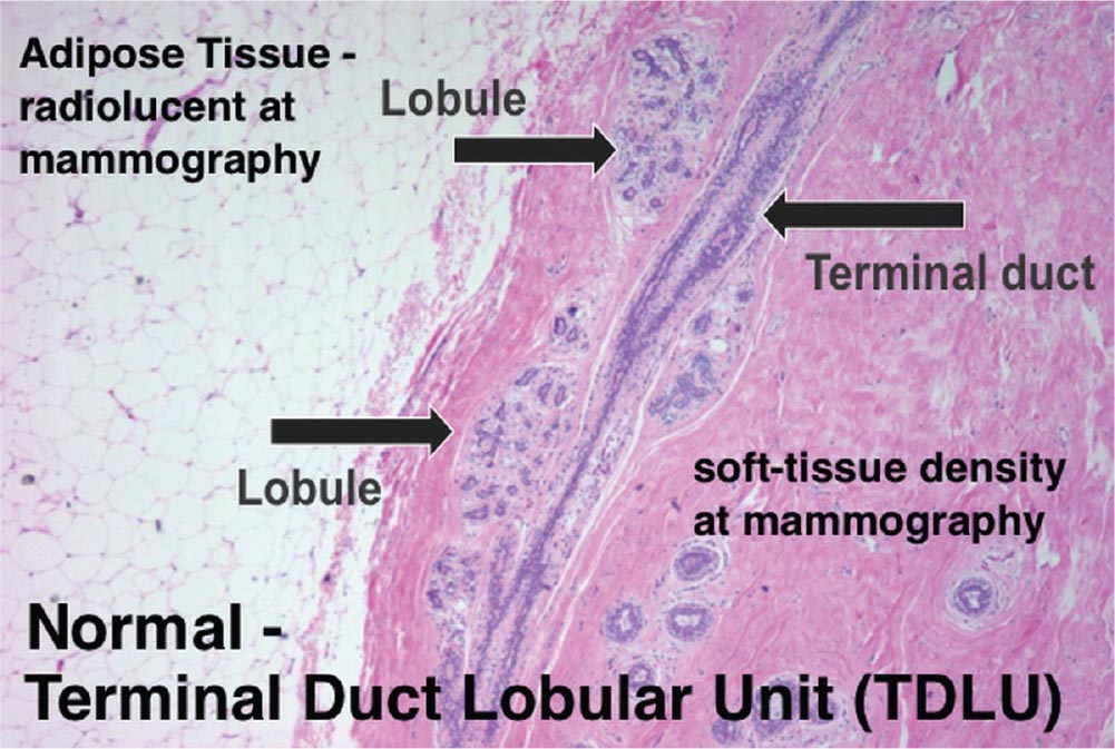
From Breast, by Thomas J. Lawton. Copyright © 2009 Cambridge University Press. Reprinted with permission.
Tips for analyzing these findings are the following:
1. In order to see “structures” on a plain film, the adjoining tissues must be of a different densities. Margins of masses and normal anatomy, such as ducts and vessels, are silhouetted by surrounding tissues of similar density. (See Fig 1–1 for histology image of a normal terminal ductal-lobular unit). This principle is key when one is analyzing margins of a suspected mass versus asymmetries.
2. Additionally, this principle is important when assessing for a fat-containing mass versus summation tissues. Note that if the margin that is being analyzed is a white line, then the margin, which is a soft-tissue density, is abutted by fat. To rule in a fat-containing mass, there must be documented volume. One must see an ovoid or round mass with a white line in two orthogonal views. Without the two planes, one cannot rule in a mass, and the white line being analyzed is a summation of normal tissues, Cooper’s ligaments.
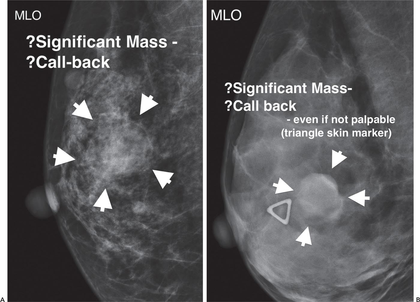
Analogously, when evaluating the mammogram for a possible significant mass, note whether the margins being connected are “lines” or a “smooth edge.” For example, Figures 1–2A and B illustrate the difference. The initial impression of Figure 1–2A at the “10,000 foot level” when images first appear is a possible mass in the superior mid-breast. However, the arrows demarcate margins that are white lines. Hence, either this is a fat-containing mass, which by definition is not significant, or summation tissues. So this is not a significant callback case. In contrast, the margins of Figure 1–2B are composed of a smooth edge. Hence the internal contents of this mass are soft tissue in density, as are the margins. This is a significant mass to callback from screening. This was a benign simple cyst.
QUESTION 4. The distinction between a focal asymmetry and a mass is a key branch point in the analysis of a mammogram since the former represents normal tissues and the latter may be malignant. What three objective criteria must one analyze?1
ANSWER 4:
1. Shape
Stay updated, free articles. Join our Telegram channel

Full access? Get Clinical Tree


