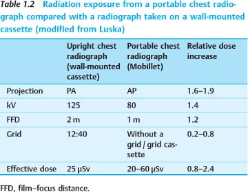
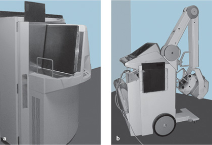
Basic Principles: Radiologic Techniques and Radiation Safety 1 Radiologic Techniques in the Intensive Care Unit Radiation Exposure and Radiation Safety Communication, Reporting of Findings, and Teleradiology Radiologic examinations in the intensive care unit (ICU) are most commonly performed at the bedside. They consist mainly of portable chest radiographs, followed by ultrasound scans. Projection radiographs of the skeleton are only rarely obtained. Much as in other clinical settings, computed tomography (CT) has assumed an expanding role in the ICU. CT scans are obtained at an increasingly early stage for addressing diagnostic problems of the chest and abdomen. This relates to the generally higher diagnostic efficiency of CT over other modalities, the capabilities of modern scanners in the detection of vascular pathology (pulmonary embolism, intestinal ischemia), and the use of CT for image-guided interventions (e. g., abscess drainage). The following typical problems are encountered in the diagnostic imaging of ICU patients: The following basic radiological equipment should be available in the ICU: The number of radiography machines and the scope of accessory equipment will depend on the number of ICU beds and on special hygienic requirements. Larger units and suites should be equipped with their own film processor or digital reader device. A portable fluoroscopy machine with its own table should be available in a separate examination room. Portable CT scanners have been tested under study conditions but have not come into practical use due to technical limitations. Conventional film–screen radiography has been replaced by digital radiography in almost all ICUs and is mentioned here only for completeness. A conventional cassette contains the x-ray film and a pair of intensifying screens placed at the front and back of the cassette. These film–screen combinations are characterized by their spatial resolution and dose requirement, which determine the speed rating of the system (50, 100, 200, 400, 800, or 1600). Faster systems require a lower radiation dose but also sacrifice some degree of spatial resolution. Film– screen combinations with a speed rating of 400 (“400 systems”) are used for radiographs in ICU patients and for most applications in emergency medicine, while 100 or 200 systems are used only in the evaluation of limb injuries (traumatology). Imaging with a scatter-reduction grid provides higher image quality than filming without a grid. While the use of a grid requires a higher exposure level (dose is generally increased by a “grid factor” of 2–3), this can be partially offset by using a higher tube voltage (120 kV instead of 80 kV for chest films). Digital radiography has become the mainstay for image acquisition and documentation in ICUs and emergency rooms owing to its technical advantages. The advantages of digital technology relate to organizational aspects (digital data format with capabilities for data transfer and image distribution to multiple users) and its lack of sensitivity to underpenetration and poor contrast due to exposure errors. These advantages are based on the digital data format, image processing including automated signal normalization, and the greater dynamic range of digital detectors compared with x-ray film. Computed radiography (CR), or storage phosphor radiography, is the current standard in intensive care radiology. Mobile flat-panel or direct radiography (DR) systems have recently become available in the 35 × 43 cm format. The advantage of DR is the greater dose efficiency of the system, which permits ca. 50% dose reduction during the examination. Computed radiography. CR (Fig. 1.1a) is comparable in its handling to a daylight system. It is a cassette-based system that requires a special reader device. The detector consists of a photostimulable storage phosphor plate housed in an aluminum cassette. After the plate has been exposed, the cassette is placed into the reader. The radiographic image can either be printed on film (hard copy) or displayed on a video monitor (soft copy). Direct detector units. Direct detector units (Fig. 1.1b) consist of the imaging cassette, which contains the detector and is hardwired to the readout unit by an electric cable. This means that the technician must take the readout unit to the patient’s bedside along with the rest of the system, which will naturally affect workflow and organizational details. The advantage of DR over CR is its higher dose efficiency, which allows for a significant dose reduction (30–50%, depending on the patient’s condition). Another advantage is the direct availability of the image on the readout unit, which provides immediate feedback on imaging parameters. Fig. 1.1 a, b Equipment for digital radiography. a Computed radiography. b Flat-panel system for direct radiography. Issues of medical radiation safety are rarely a priority concern in emergency and ICU patients, even though the patients may be exposed to considerable dose levels. One problem is that trauma patients in particular must often undergo comprehensive imaging protocols that involve high individual doses during the acute phase of treatment. Another problem is that even low individual doses per examination may well produce a significant cumulative exposure when continued over a period of weeks. The supine anteroposterior (AP) chest radiograph is the most common imaging examination in the ICU, especially in patients on long-term ventilation. More than 100 radiographs may be taken during a prolonged stay in the ICU. While various dose values have been reported in the literature, the effective dose in all cases was less than 0.2 mSv per radiograph. CT examinations expose patients to significantly higher effective dose values than chest radiographs (Table 1.1). The values listed in the table are only approximations, however, as the effective dose depends strongly on equipment and examination parameters. A portable chest radiograph generally exposes the patient to more radiation than a film taken on a wall-mounted cassette holder, depending on the selected parameters (Table 1.2). This is due mainly to the shorter film–focus distance (FFD) at the bedside and the use of an AP rather than posteroanterior (PA) projection (increasing the effective dose to females by a factor of 1.9, to males by a factor of 1.6). The dose increase associated with the use of a scatter-reduction grid (grid cassettes) can be partially offset by a higher kilovoltage setting. The risk coefficients defined by the ICRP (International Commission on Radiological Protection) in 1991 are useful for estimating the bioeffects of radiation. The likelihood that a 30-year-old individual will develop a radiation-induced malignancy is estimated at 5% per sievert(4.5%/Sv for solid cancers, 0.5%/Sv for leukemia; ICRP 60; see also Table 1.5). The latent period for developing leukemia is ca. 15 years compared with 40 years for other malignant diseases. This latent period is a particular concern in older individuals. Based on the above percentages, an ICU patient who receives 100 chest radiographs (total dose of 0.02 Sv based on single doses of 20 μSv) will have an additional 0.1% risk for developing a malignant disease (Table 1.3). Since approximately one in four people will develop a malignancy during their lifetime, the chest radiographs in the above example will increase the risk from 25% to 25.1%. Thus, even in a setting of long-term intensive care involving multiple radiographic examinations, the patient will not face a significant additional cancer risk, especially when we consider the severity of the condition for which the patient is receiving intensive care. Acute illness or injury in a pregnant woman, while rare, is a challenging management problem. One aspect of the problem concerns the use of roentgen rays and the resulting radiation exposure to the embryo or fetus. Antenatal exposure to ionizing radiation can potentially lead to two types of pathology: malignancies and malformations.
Radiologic Techniques in the Intensive Care Unit
C. Schaefer-Prokop
 Patients have limited ability to cooperate with the examiner.
 Imaging conditions are more difficult than in the radiology department (chest imaged in a supine or sitting position, limited access for ultrasound scans, etc.).
 Radiographic interpretation is often hampered by superimposed foreign materials (dressings, metal implants, catheters, tubes, wires).
 Radiographic equipment is frequently limited (portable radiography machine), and images are obtained without automatic exposure control.
 If the patient is taken to the radiology department (e. g., for CT, MRI, DSA), the gain in diagnostic information must be weighed against the increased risk of transporting the patient.
Radiographic Equipment
 a portable radiography machine
 storage phosphor cassettes, or cassette-based direct detectors with a 35 × 43 cm format—may be used as a grid-film cassette or with a Bucky device for inserting standard film cassettes
 radiation protection aprons (lead equivalency of 0.25–0.5 mm) and protective gloves
 portable radiation screens for some applications
 viewboxes or video monitors for viewing images (large enough for viewing and comparing two or three large-format films)
 ultrasound scanner with documentation system
Conventional Film–Screen Radiography
Digital Radiography
Radiation Exposure and Radiation Safety
A. Stadler
Radiation Exposure to Patients and Staff
Assessment of Patient Risk
Radiation Exposure during Pregnancy
Type of examination | Typical effective dose |
PA chest radiograph | 0.025 mSv |
AP chest radiograph | 0.06 mSv |
AP abdominal radiograph | 1 mSv |
Cranial CT | 2.3 mSv |
Thoracic CT | 8 mSv |
Abdominal/pelvic CT | 10 mSv |
 Digital radiography has almost completely replaced conventional film–screen radiography in ICUs.
 Computed radiography is the standard in ICUs and emergency rooms; digital radiography machines have also become available in recent years.
 Even numerous radiographic examinations do not pose a significant additional cancer risk in ICU patients.
 A portable chest radiograph exposes the patient to more radiation than a film taken on a wall-mounted cassette holder.