KEY FACTS
Terminology
- •
Saccular outpouching from herniation of bladder mucosa and submucosa through muscular wall of bladder
Imaging
- •
Most commonly near ureterovesical junction
- •
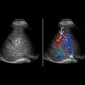
Anechoic outpouching from bladder with narrow or wide neck, may empty with micturition
- •
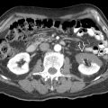
CT/MR: Fluid attenuation outpouching from bladder
- •
Usually fills with contrast on excretory postcontrast phase
Top Differential Diagnoses
- •
Urachus
- •
Everted ureterocele
- •
Paraovarian cysts in female
- •
Pelvic cysts in male
Pathology
- •
Acquired: Most common secondary to chronic bladder outlet obstruction (60%)
- •
Congenital: Hutch diverticulum (40%)
- •
Vesicoureteral reflux
Clinical Issues
- •
Narrow-neck diverticula: Urinary stasis → complications such as infection, stone, and ureteral obstruction
- •
Secondary inflammation predisposes to development of carcinoma within diverticulum
- •
Complications
- ○
Carcinoma
- ○
Vesicoureteral reflux
- ○
Ureteral obstruction
- ○
Scanning Tips
- •
Look for debris, calculi, or solid masses in diverticula, which can indicate complication
- •
Color Doppler may show jet to and from diverticulum to bladder; do not mistake for ureteral jet
 arising from the lateral bladder wall, due to herniation of the mucosa and submucosa through the muscular wall.
 with narrow necks
  . One of the diverticula shows a urinary jet as Doppler signal into the diverticulum
  .
.
Stay updated, free articles. Join our Telegram channel

Full access? Get Clinical Tree


