, Lawrence A. Zumo2 and Valerie Sim3
(1)
Parkinson’s Clinic of Eastern Toronto, Toronto, ON, Canada
(2)
Silver Spring, Cheverly, MD, USA
(3)
Centre for Prions and Protein Folding Diseases, University of Alberta, Edmonton, AB, Canada
Abstract
Imaging of the blood vessel lumen can be best achieved by injecting a dye into the vessel and then imaging the dye with Xray, CT, or MR technologies, but a lot of information can be gleaned by measuring flow through the vessels with ultrasound. Because of the increased risk of stroke with conventional angiography, CT angiography or MR angiography is often the first choice of investigation for imaging intracranial arteries, but doppler ultrasound is still frequently used to assess the carotid arteries. Interpreting the results from any modality requires a good knowledge of vessel anatomy. In this chapter we present a quick review of the anatomy of the circle of Willis and provide case examples of vessel stenosis and aneurysms using MRA, CTA, and doppler ultrasound.
Case 11.1 Anatomical Review
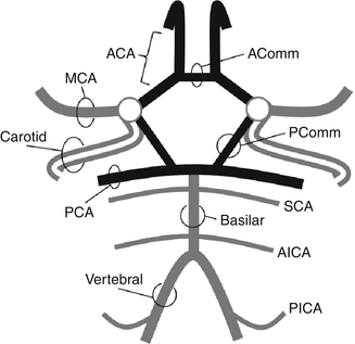
Figure 11.1
Diagram of the Circle of Willis and major intracranial blood vessels. ACA anterior cerebral artery, AComm anterior communicating artery, MCA middle cerebral artery, PComm posterior communicating artery, PCA posterior cerebral artery, SCA superior cerebellar artery, AICA anterior inferior cerebellar artery, PICA posterior inferior cerebellar artery
Case 11.2 Aneurysm (MRA)
< div class='tao-gold-member'>
Only gold members can continue reading. Log In or Register to continue
Related posts:


Stay updated, free articles. Join our Telegram channel

Full access? Get Clinical Tree


