BONY ORBIT AND EXTRACONAL COMPARTMENT: TUMORS
KEY POINTS
- Computed tomography and magnetic resonance imaging are in the critical pathway for evaluating the signs and/or symptoms that are related to an extraconal-origin orbital neoplasm.
- Computed tomography and magnetic resonance imaging are extremely useful for narrowing the differential diagnosis of disease that may arise outside the muscle cone but has limits in differentiating benign tumors or low-grade malignant tumors and sometimes vascular malformations.
- Imaging with magnetic resonance or computed tomography will help establish whether the disease process is localized or possibly related to systemic disease.
- If a lesion is not to be biopsied or removed, magnetic resonance or computed tomography should be used to establish whether its rate of growth is consistent with a benign or malignant process.
Any orbital mass or disease process may be approached by first establishing whether it is preseptal (Chapters 70 and 71) or postseptal (Chapters 57–60, 62, and 64). If postseptal, it should be established as a process arising primarily intraconally (Chapters 57–60), extraconally (Chapters 62 and 64), or transcompartmentally. If intraconal, it should be decided whether the mass is of optic nerve/sheath origin or arises from some other structure comprising or within the muscle cone. This chapter considers extraconal tumors and those arising extraconally and spreading across the orbital compartments and related spaces.
Extraconal orbital lesions arise within the postseptal extraconal space, subperiosteal space, and within bone. They may also be a manifestation of spread along neurovascular bundles that course within extraconal fat and/or travel subperiosteally at various points along their periorbital distribution—the prime examples being the supraorbital and infraorbital neurovascular bundles. Transcompartmental lesions will involve one or more of these spaces and may spread to involve the muscle cone and its central surgical space.
The spread patterns of these tumors are discussed in the following Pathophysiology and Patterns of Disease section.
ANATOMIC AND DEVELOPMENTAL CONSIDERATIONS
Applied Anatomy
The relevant anatomy of the bony orbit, muscle cone, and related neurovascular structures is discussed in detail in Chapter 44. In particular, the anatomy of the orbital periosteum, orbital septum, and attachments of the muscle cone at the orbital apex should be reviewed. The subperiosteal space within the extraconal, postseptal compartment lies between cortical bone and its adjacent periosteum. The postseptal extraconal space lies between the periosteum and muscle cone and contains fat and small nerves and vessels.
IMAGING APPROACH
Techniques and Relevant Aspects
The orbit is studied with computed tomography (CT) and magnetic resonance (MR) techniques described in detail in Chapters 44 and 45. Specific CT protocols by indications are detailed in Appendix A. Specific MR protocols by indications are outlined in Appendix B. Almost all studies to investigate orbital tumors are done with contrast except if the lesion is of known fibro-osseous origin.
Pros and Cons
Disease of extraconal orbital spaces is studied primarily with CT and magnetic resonance imaging (MRI). CT may be done first since it is more definitive in its rendering of bone detail, which is often pivotal in the diagnosis. MRI is likely to be more sensitive than CT to marrow space changes within bone, perineural spread, and possibly related intracranial abnormalities such as early dural or pia-arachnoid changes associated with these tumors.
CT may also be done first for an emergent evaluation of possible associated tension orbit. It is also a first choice in patients too sick or for some other reason unable to complete a very high quality, free of motion artifact, MRI examination. CT is a reasonable secondary screening tool for identifying intracranial extension of disease and intracranial complications.
SPECIFIC DISEASE/CONDITION
Benign and Malignant Tumors
Etiology
Benign tumors of the extraconal spaces are rare except for those that arise in bone or the sinuses and nasal cavity (Chapters 89–92). Those in bone are predominantly fibro-osseous tumors (Chapters 40 and 91) and meningiomas (Chapter 31). Neurogenic tumors (Chapter 30) track along nerves in the extraconal fat.1–3
Malignant tumors of the extraconal spaces are rare except for those that arise in bone, especially the greater wing of the sphenoid or the sinuses and nasal cavity (Chapters 89–92). Those in bone are predominantly metastatic tumors or manifestations of lymphoma, leukemia, or plasma cell dyscrasias (Chapters 27, 28, and 42).4,5
Prevalence and Epidemiology
These lesions are spontaneous occurrences in children and adults. Neurogenic tumors associated with neurofibromatosis type 1 often spread in the extraconal fat (Chapter 29).
Clinical Presentation
Benign tumors may present with painless proptosis. There may eventually be visual loss, diplopia, and/or other manifestations of ocular dysmotility.
Pathophysiology and Patterns of Disease
These tumors grow in a fashion typical of benign and malignant tumors as described in Chapter 21.
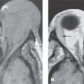
The extraconal space is commonly involved in orbital pathology, but it is most often not the site of disease origin. Superior and medial wall or orbital floor lesions usually arise from sinonasal pathology. Superior and lateral lesions are often of lacrimal gland origin (Chapter 66). Practically speaking, the involvement of the extraconal fat is usually due to pathology involving at least one other adjacent space and/or bone of the extraconal compartment (Fig. 64.1). These masses may have an ill-defined infiltrating morphology. When an infiltrating transcompartmental process appears to be centered in the extraconal space, the pattern strongly suggests lymphoma or other malignancy (Figs. 64.2 and 64.3) versus idiopathic orbital inflammatory syndrome (IOIS) or one of the granulomatoses or histiocytoses as an etiology.

FIGURE 64.1. Four patients with proptosis. A: Computed tomography (CT) of a patient presenting with slow onset of proptosis due to fibrous dysplasia of the greater wing of the sphenoid bone (arrows). B: CT scan of a meningioma centered in the greater wing of the sphenoid bone (black arrow) and showing a very destructive pattern with an osteoid-appearing matrix (arrowheads). Metastases and osteosarcoma were considered in the differential diagnosis. C: CT showing sclerosis of the greater wing of the sphenoid on the right (arrow) compared to that on the left (arrowhead). D: Soft tissue window of the image in (C) shows extension of a mass into the orbital soft tissues (arrowhead) and intracranially (arrows) due to metastatic Ewing sarcoma.
Perineural spread of cancer along the first and second divisions of the trigeminal nerve often due to skin and other cancers (Chapters 21, 22, and 24) or neurogenic tumors (Chapter 29) may spread along vessels in the extraconal space (Figs. 64.4 and 64.5).
Disorders arising in the extraocular muscles (Chapter 60) frequently involve the extraconal fat. Focal enlargement of an extraocular muscle extending into the extraconal fat is likely due to metastasis or lymphoma (Fig. 64.3).
Extraconal subperiosteal tumor elevates the periosteum and creates a fusiform mass lacking abrupt transition zones with adjacent normal periosteum (Figs. 64.1A, 64.3, and 64.4). This is analogous to a pleural or extrapleural mass on chest imaging. An associated focal tumor is usually evident. When an apparently subperiosteal mass is not associated with contiguous bone or sinus abnormalities, an inflammatory or neoplastic infiltrative process such as myeloproliferative and lymphoproliferative diseases (Fig. 64.2C and Chapter 27), pseudotumor (Chapter 57), or a granulomatosis actually centered in the extraconal fat should be considered. When subperiosteal lesions are identified, it is critical to determine whether the dura is also thickened, displaced, and/or penetrated by the pathologic process (Fig. 64.1C). In the medial orbit, subperiosteal tumors are of sinonasal origin or those of bone (Fig. 64.6 and 64.7). Subperiosteal masses along the lateral orbital wall and greater wing of the sphenoid are usually associated with metastatic tumor (Fig. 64.1A and Chapter 42), leukemia, lymphoma or plasmacytoma of the diplopic space (Chapter 27), or transdiploic spread of meningiomas (Fig. 64.1B and Chapter 31).

FIGURE 64.2. Two patients presenting with proptosis due to lymphoma. A, B: Patient 1. In (A), contrast-enhanced T1-weighted (T1W) magnetic resonance shows an infiltrating extraconal mass (arrows) with no bone destruction. In (B), another section from the same sequence shows infiltration along the course of V2 in the inferior orbital fissure and pterygomaxillary fossa region (arrowhead) as well as along the foramen rotundum (arrow). (NOTE: Neurotropic behavior of this lesion combined with the extraconal involvement strongly suggests the correct diagnosis of lymphoma.) C: Patient 2, presenting with orbital lymphoma. Contrast-enhanced T1W fat-suppressed imaging showing extraconal infiltration in both orbits (arrows) and a suggestion of leptomeningeal involvement intracranially (arrowhead). (NOTE: The bilateral disease and intracranial disease suggested a systemic infiltrating tumor; this pattern strongly suggests the correct diagnosis of lymphoma.)
Stay updated, free articles. Join our Telegram channel

Full access? Get Clinical Tree


