Both Column Acetabular Fracture
Brian P. Milam
Daniel B. Nissman
CLINICAL HISTORY
20-year-old female who was transported to the ED by EMS after a restrained motor vehicle collision. She is complaining of left leg and hip pain.
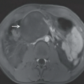
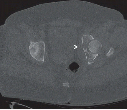 FIGURE 36A |
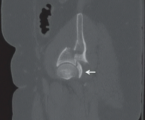 FIGURE 36B |
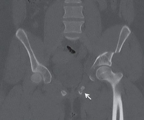 FIGURE 36C |
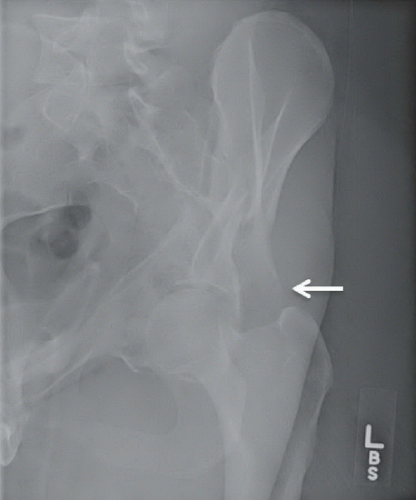 FIGURE 36D |
FINDINGS
Axial CT image (Fig. 36A) depicts a comminuted fracture with a dominant coronal plane fracture line (arrow) dividing the acetabulum into anterior and posterior portions. Sagittal CT image (Fig. 36B) demonstrates a comminuted fracture involving the acetabular roof; there is posterolateral displacement of the ilium relative to the acetabulum and a nondisplaced posterior wall fracture fragment (arrow). Coronal CT image (Fig. 36C) shows a minimally displaced inferior pubic ramus fracture (arrow) and the medially impacted comminuted left acetabular fracture. An obturator oblique radiograph (Fig. 36D) shows a comminuted left acetabular fracture with disruption of both the iliopectineal and ilioischial lines. The unsupported left ilium fragment comes to a point inferiorly and laterally, the “spur” sign (arrow).
DIFFERENTIAL DIAGNOSIS
Anterior column acetabular fracture, posterior column acetabular fracture, T-type acetabular fracture, both column acetabular fracture.
DIAGNOSIS
Both column acetabular fracture.
DISCUSSION
Acetabular fractures are most often seen in patients in their third to fifth decades that have experienced high-energy trauma.1 They can also occur in patients of advanced age owing to low-energy trauma, such as a fall; however, this is seen much less often and is caused by osteoporotic changes. Because of the many types of acetabular fracture patterns with disparate management implications, the ability to accurately describe an acetabular fracture is essential. Both column fractures are most often associated with high-energy trauma, regardless of patient age, and are always unstable. Both column fractures are the most common type of acetabular fracture, making up approximately 27% of all diagnosed acetabular fractures.1
Related posts:


Stay updated, free articles. Join our Telegram channel

Full access? Get Clinical Tree


