GROSS ANATOMY
Supratentorial Structures
- •
Gyri : Complex convolutions of brain cortex; hypoechoic on US
- •
Sulci (fissure): Cerebral spinal fluid (CSF)-filled grooves or clefts that separate gyri; echogenic on US
- ○
Sulci separate gyri, fissures separate hemispheres/lobes
- ○
- •
Frontal lobe
- ○
Central sulcus separates frontal, parietal lobes
- ○
Precentral gyrus contains primary motor cortex
- ○
- •
Parietal lobe
- ○
Posterior to central sulcus
- ○
Separated from occipital lobe by parietooccipital sulcus (medial surface)
- ○
Postcentral gyrus: Primary somatosensory cortex
- ○
- •
Occipital lobe
- ○
Posterior to parietooccipital sulcus
- ○
Primary visual cortex
- ○
- •
Temporal lobe
- ○
Inferior to sylvian fissure
- ○
Primary auditory cortex
- ○
Middle temporal gyrus: Connects with auditory, somatosensory, visual association pathways
- ○
- •
Insula
- ○
Lies deep in floor of sylvian fissure, overlapped by portions of frontal, temporal, & parietal lobes, called the opercula
- ○
- •
Limbicsystem
- ○
Includes amygdala, hippocampus, thalamus, hypothalamus, basal ganglia, & cingulate gyrus
- –
Cingulate gyrus is directly above & parallels the corpus callosum
- –
- ○
Important role in emotion, behavior, & long-term memory
- ○
- •
Corpus callosum is white matter tract, which links cerebral hemispheres
- ○
Genu is front-most portion; the body arches over the cavum septi pellucidi, ending at the splenium above vermis
- –
Rostrum is short posterior extension from inferior portion of genu
- –
- ○
- •
Basal ganglia are paired deep gray matter structures, including the caudatenuclei
- •
Thalami are paired, large nuclear complexes, which act as relay stations for most sensory pathways
Posterior Fossa (Infratentorial) Structures
- •
Protected space surrounded by calvarium
- ○
Bounded by tentorium cerebelli superiorly (an extension of the dura mater similar to falx) & foramen magnum inferiorly (where spinal cord exits skull)
- ○
- •
Posterior fossa contents
- ○
Brainstem (midbrain, pons, & medulla oblongata) anteriorly, cerebellum posteriorly
- ○
Cerebral aqueduct & 4th ventricle
- ○
CSF cisterns containing cranial nerves, vertebrobasilar arterial system, & veins
- ○
- •
Cerebellum
- ○
Integrates coordination & fine-tuning of movement & regulation of muscle tone
- ○
2 hemispheres & midline vermis
- –
Vermis divided into lobes & lobules by multiple fissures
- □
Appears highly echogenic on US
- □
- –
Hemispheres have thin, curved gyri called folia
- –
- ○
Connected to brainstem by 3 paired peduncles
- ○
- •
Brainstem
- ○
3 anatomic divisions
- –
Midbrain (mesencephalon) : Upper brainstem; connects pons & cerebellum with forebrain
- –
Pons : Bulbous midportion of brainstem; relays information from brain to cerebellum
- –
Medulla : Caudal (inferior) brainstem; relays information from spinal cord to brain
- –
- ○
Ventricular System & Subarachnoid Space
- •
Cerebral ventricles consist of paired, lateral, midline 3rd & 4th ventricles
- •
Communicate with each other as well as central canal of spinal cord & subarachnoid space
- •
Direction of CSF flow
- ○
Lateral ventricles → foramen of Monro → 3rd ventricle → cerebral aqueduct (of Sylvius) → 4th ventricle → foramina of Luschka & Magendie → subarachnoid space
- ○
Bulk of CSF resorption through arachnoid granulations in superior sagittal sinus
- ○
- •
Lateral ventricles
- ○
Paired, C-shaped structures, which arch around/above thalami
- ○
Each has body, atrium, 3 horns (frontal, temporal, & occipital)
- –
Occipital horn typically largest
- –
Asymmetry is common, often L > R
- –
Sizes change with maturity, more prominent in preterm infants
- –
- ○
Atrium/trigone : Confluence of horns
- –
Contains glomus (thickened portion) of choroid plexus
- –
- ○
Lateral ventricles communicate with each other & 3rd ventricle via Y-shaped foramen of Monro
- ○
- •
3rd ventricle
- ○
Thin, usually slit-like, between thalami
- –
May not see fluid, just bright echogenic line on US
- –
- ○
80% have central adhesion between thalami ( massa intermedia )
- ○
There are 4 small recesses: 2 projecting anteriorly (optic & infundibular recesses) & 2 projecting posteriorly (suprapineal & pineal)
- ○
Communicates with 4th ventricle via cerebral aqueduct (of Sylvius), passing through dorsal midbrain
- ○
- •
4th ventricle
- ○
Infratentorial, diamond-shaped cavity between brainstem & vermis
- ○
Fastigial point : Blind-ending, dorsally pointed midline outpouching from body of 4th ventricle
- –
Important marker for true midline vermian plane on US
- –
- ○
CSF exits 4th ventricle into subarachnoid space via foramina of Magendie (midline) & Luschka (lateral)
- ○
Inferiorly communicates with central canal of spinal cord
- ○
- •
Choroid plexus
- ○
Produces CSF
- ○
Glomus (enlargement of choroid plexus in atrium) thickest area
- ○
Tapers & extends anteriorly to foramen of Monro & roof of 3rd & 4th ventricles
- ○
Tapers laterally into roof of temporal horns
- ○
Never extends into frontal or occipital horns
- ○
- •
Subarachnoid space/cisterns
- ○
CSF-containing spaces around brain
- ○
Numerous trabeculae, septa, membranes cross subarachnoid space & create smaller compartments termed cisterns
- –
Cisterna magna is large cistern in posterior fossa
- –
- ○
All cisterns communicate with each other & with ventricular system
- ○
- •
Midline cystic structures (normal variants)
- ○
Cavum septi pellucidi
- –
Anterior to foramen of Monro, between anterior horns of lateral ventricles
- –
85% closed by 3-6 months after birth, but some remain open into adulthood
- □
Once closed called septum pellucidum
- □
- –
- ○
Cavum vergae
- –
Posterior to foramen of Monro, interposed between bodies of lateral ventricles
- –
Posterior extension of cavum septi pellucidi ( cavum septi pellucidi et vergae )
- –
Begins to close from posterior to anterior from 6-months gestation; 97% closed by full term
- –
- ○
Cavum velum interpositum
- –
Potential space, which may accumulate CSF, above choroid in roof of 3rd ventricle & below fornices
- –
Typically seen in premature infants
- –
- ○
ANATOMY IMAGING ISSUES
Imaging Approaches
- •
All scanning should be performed keeping the exposure as low as reasonably achievable, the ALARA principle
- •
Scans should be performed using a small footprint, high-frequency transducer, which allows sufficient penetration to see deep structures
- ○
Use linear high-frequency transducer for evaluating superficial structures, such as extraaxial fluid spaces (e.g., subdural hematoma) or superior sagittal sinus (e.g., thrombosis)
- ○
- •
Use Doppler (color, spectral, &/or power) as needed to evaluate vasculature structures
- ○
Always use to confirm an anechoic structure is truly a cyst & not a vascular malformation
- ○
- •
Anterior fontanelle most commonly used approach
- •
Coronal scans
- ○
Complete sweep from front to back documenting key landmarks
- –
Begin anterior to frontal horns & extend to posterior to occipital horns, ensuring entire brain has been covered
- □
May need to tilt transducer laterally to include superficial peripheral surfaces
- □
- –
Adjust transducer position as needed to keep image symmetric side to side
- –
Adjust depth to include posterior fossa, including cerebellar hemispheres & cisterna magna
- –
- ○
Symmetrical structures (from anterior to posterior) include frontal horns, bodies, trigones & occipital horns of lateral ventricles; caudate nuclei & thalami
- –
Foramina of Monro seen extending inferomedially into 3rd ventricle creating a Y shape
- □
Just posterior to this will be 3-dot sign : Choroid plexus on floor of lateral ventricles & roof of 3rd ventricle
- □
Choroid plexus does not extend anterior to this point; echogenic material anterior to this would represent hemorrhage
- □
- –
- ○
Midline structures include interhemispheric fissure, genu & body of corpus callosum, cavum septi pellucidi, 3rd ventricle, vermis, & 4th ventricle
- ○
- •
Sagittal scans
- ○
Midline scan: Best view for corpus callosum, cerebellar vermis, & 4th ventricle
- ○
Sweep side to side from this position documenting key areas
- –
Caudothalamic groove : Site of germinal matrix (highly vascular area from which cells migrate during brain development)
- □
Most common site of hemorrhage in premature infants
- □
- –
Size of lateral ventricle
- –
Far lateral to assess degree of sulcal development
- –
- ○
- •
Posterior fontanelle
- ○
Best view to evaluate occipital horns for intraventricular hemorrhage
- –
Can misinterpret clot adherent to choroid plexus from anterior fontanelle approach alone
- –
Use color Doppler to confirm flow in choroid plexus
- –
- ○
- •
Mastoid fontanelle
- ○
Located at junction of squamosal, lambdoidal, occipital sutures
- ○
Transducer placed about 1 cm behind helix of ear & 1 cm above tragus
- ○
Allows assessment of brainstem & posterior fossa
- ○
Best view for 4th ventricle, posterior cerebellar vermis, cerebellar hemispheres, & cisterna magna
- ○
- •
Transtemporal
- ○
Temporal bone anterior to ear is thin, allowing imaging of brainstem even after sutural closure
- ○
Best view for cerebral peduncles & 3rd ventricle
- ○
Imaging Pitfalls
- •
Need to understand the normal development & changing appearance of the brain as it matures
- •
Must know not only age in days/weeks of infant but gestational age at birth
- ○
Normal gyral pattern in 26-week preterm infant would be abnormal in term infant
- ○
- •
Slit-like lateral ventricles common in infants, not to be mistaken for cerebral edema
- •
Glomus of choroid plexus can be bulbous & irregular, not to be mistaken for blood clot
- ○
Evaluate with color Doppler & posterior fontanelle view
- ○
- •
Echogenic material in frontal or occipital horns is clot; choroid does not extend into these horns
BRAIN


VENTRICULAR SYSTEM

STANDARD US PLANES VIA ANTERIOR FONTANELLE


CORONAL US VIA ANTERIOR FONTANELLE


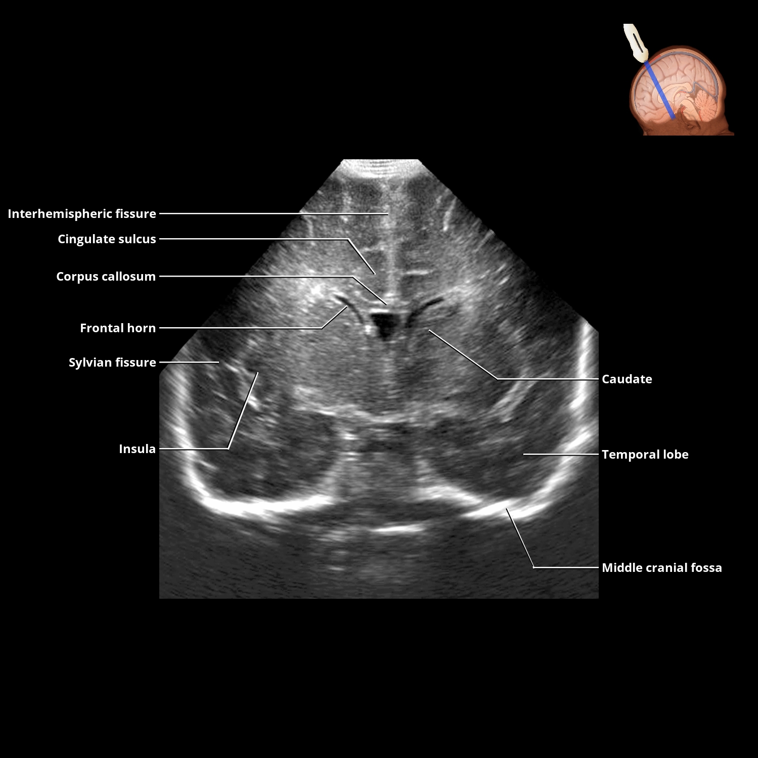
CORONAL US VIA ANTERIOR FONTANELLE


CORONAL US VIA ANTERIOR FONTANELLE


SAGITTAL US VIA ANTERIOR FONTANELLE


SAGITTAL US VIA ANTERIOR FONTANELLE