KEY FACTS
Terminology
- •
Placenta detachment
- ○
Most often partial and can see hematoma
- ○
- •
Categorized by location of abruption
Imaging
- •
Marginal abruption (most common)
- ○
Hemorrhage from edge of placenta
- ○
- •
Retroplacental abruption (2nd most common)
- ○
Hematoma between placenta and uterus
- ○
May be confused for placentomegaly when acute
- ○
- •
Preplacental abruption (rare)
- ○
Hematoma on fetal surface of placenta
- ○
- •
Hematoma appearance depends on age of blood
- ○
Acute hematoma: Often isoechoic to placenta
- ○
Subacute hematoma: Hypoechoic to placenta
- ○
Resolving/chronic hematoma: Sonolucent
- ○
Top Differential Diagnoses
- •
Leiomyoma: Will see flow on color Doppler
- •
Focal myometrial contraction: Changes during scan
Clinical Issues
- •
Abruption is clinical diagnosis: Only 1/3 will have findings
- •
Risk factors: Trauma (particularly MVA), gestational hypertension, smoking, placenta previa, prior abruption
Scanning Tips
- •
Use color Doppler to differentiate isoechoic clot (no flow) from placenta
- •
Scan entire uterus
- ○
Hematoma may be distant from placenta edge
- ○
- •
Look for abruption and previa in all 2nd- and 3rd-trimester cases with vaginal bleeding or tender uterus
- •
Evaluate fetal heart rate early if new diagnosis of abruption
- ○
Stop scanning and call for help if bradycardia
- ○
Emergency cesarean warranted if viable fetus
- ○
 occurs at the placental edge. Retro-PA
  occurs between the placenta and uterine wall, and pre-PA
  occurs in front of the placenta, between the placenta and membranes.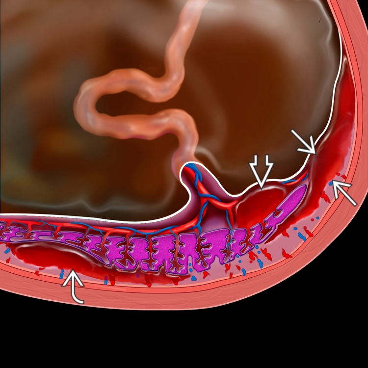
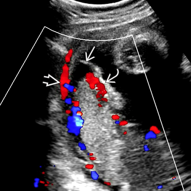
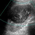
 has been lifted off the uterine wall
  . A small sonolucent hematoma is seen
  , suggesting the abruption is old. Color Doppler shows flow in the placenta and uterus but not in the clot.