LEARNING OBJECTIVES
1. Overview of risk factors associated with breast cancer risk
• Chemoprevention
2. Screening mammography
• Randomized controlled trials
• Controversies
• Screening guidelines
3. Mammography Quality Standards Act
4. American College of Radiology: Mammography Accreditation Program
5. Overview of treatment options
• Lumpectomy, mastectomy
• Sentinel lymph node biopsy versus axillary lymph node dissection
• Radiation therapy
Accelerated partial breast radiation
• Chemotherapy
Use of Oncotype DX
Neoadjuvant therapy
• Chemoprevention
6. Breast cancer staging
• TNM
• Genetic profiling of breast tumors
7. Considerations when reviewing patient images in breast imaging
Breast cancer is a common, life-threatening disease. If skin cancers are excluded, breast cancer is the most frequently diagnosed malignancy and the second leading cause of cancer mortality among women second only to lung cancer (1,2). Women have a 12.5% (1 in 8 women) lifetime risk of developing breast cancer by age 85 (3,4). It is estimated that 232,670 women and 2,360 men will be diagnosed with invasive breast cancer in the United States in 2014. An additional 62,570 women are estimated to be diagnosed with in situ breast cancer, 85% of which will be ductal. It is estimated that 40,430 breast cancer–related deaths will occur in the United States in 2014 (40,000 in women, 430 in men) (1,2). Breast cancer–related deaths have been progressively decreasing since 1989 such that approximately 30% fewer women are dying from it annually (5). A close to 7% decrease in the incidence rate of breast cancer reported from 2002 to 2003 is attributed in part to the reduction in the use of hormone replacement therapy. No change in the incidence rate for breast cancer was reported between 2005 and 2009 (1,2).
RISK FACTORS
Many factors are reportedly associated with an increased risk of breast cancer, but only a few are considered significant. Included among these are gender, age, a personal or family history of breast cancer in first-degree relatives, prior breast biopsy with certain histological diagnoses, a history of high-dose radiation to the chest between the ages of 10 and 30, and mutations in BRCA1 and BRCA2 genes as well as several other relatively rare breast cancer susceptibility genes (2–4,6). Other factors that have been implicated include: early menarche, late menopause, late first-term pregnancy, nulliparity, and postmenopausal obesity. For these risk factors, prolonged exposure of breast tissue to estrogen is thought to play a role (2–4,6). The density of the breast parenchyma on a mammogram has also been suggested and described as an indicator for increased breast cancer risk (2–4,6–13); the extent to which this is true remains to be defined (see below) (14).
Women are about 100 times more likely to develop breast cancer than men, and the incidence of breast cancer increases as women get older (2). The risk increases rapidly in premenopausal women and more slowly in postmenopausal women. Women with a personal history of breast cancer have a three- to fourfold increase in the risk of developing a new breast cancer compared with women who have never had the disease. The risk of developing breast cancer is doubled in women with one, and increased threefold with two first-degree relatives with breast cancer (mother, sisters, or daughters), particularly if the breast cancer in the relative developed premenopausally and was bilateral or multiple. The associated risk for women who have had a father or brother with breast cancer is increased, but it is not known by exactly how much. It is important to emphasize, however, that approximately 85% of women diagnosed with breast cancer do not have a family history of the disease (3,4,6).
Women who have had a breast biopsy may also be at increased risk for the development of breast cancer (2). Benign breast disease can be subdivided into three general categories: (i) Non-proliferative lesions have little effect on the risk for breast cancer; these include fibrosis and fibrocystic changes, apocrine and squamous metaplasia, mild hyperplasia, adenosis, solitary papillomas, fat necrosis, duct ectasia, lipoma, fibroadenolipoma, neurofibroma, and vascular lesions. (ii) Proliferative lesions with no atypia are reportedly associated with an increased risk of 1.5 to 2 times that of “normal” women. These processes include ductal hyperplasia of the usual type, fibroadenoma, sclerosing adenosis, multiple papillomas, and complex sclerosing lesions. (iii) Proliferative lesions with atypia increase the risk in patients by 3.5 to 5 times that of patients who do not have these processes. Atypical ductal hyperplasia (ADH) and atypical lobular hyperplasia (ALH) are the two primary lesions in this group. The associated risk is even higher for patients with these proliferative processes and a family history of breast cancer. The significance and risk associated with more recently described pathological entities such as flat epithelial atypia, columnar cell change with atypia, and atypical papillomas are not yet adequately defined. Unlike ductal carcinoma in situ (DCIS) that is considered to be a cancer, lobular carcinoma in situ (LCIS) has traditionally been considered a marker lesion of increased risk and not a precursor. Patients with LCIS have a 7- to 11-fold increase in the risk of developing breast cancer. The risk associated with LCIS applies to both breasts, and the patients have an equal likelihood of developing invasive ductal or lobular cancers (3,4,6,15).
Two genes (BRCA1, BRCA2) have been isolated in some women with breast cancer. It is estimated that 5% to 10% of all breast cancers in the United States are hereditary, related to mutations in the BRCA1 or BRCA2 genes; these genes are more common in Jewish people of Ashkenazi descent, but can occur in anyone. Women with mutations in the BRCA1 gene are also at increased risk for developing ovarian cancer. The risk of developing breast cancer in women who carry mutations in this gene is 85% by age 70 (63% for ovarian cancer by age 70); the average risk for breast cancer in BRCA1 carriers is 55% to 65%. These patients are likely to be younger and premenopausal with multiple tumors and bilateral synchronous or metachronous lesions. The tumors in these patients are often aggressive and phenotypically triple negative (e.g., estrogen receptor, progesterone receptor, and HER2/neu negative). Women who carry mutations in the BRCA2 gene have an increased breast cancer risk of approximately 45% but are not at increased risk for ovarian cancer. Breast cancers in patients with this mutation are also more likely to develop in younger women and be bilateral. Hereditary male breast cancer is associated with mutations in the BRCA2 gene but not with BRCA1. There are some genetic diseases (Li-Fraumeni syndrome, Cowden disease, Bannayan–Riley–Ruvalcaba syndrome, and ataxia-telangiectasia) associated with breast cancer susceptibility genes and an increased risk of breast cancer among affected patients (2–4,6). Genetic testing for the BRCA1 and BRCA2 mutations as well as other genes (PTEN or TP53) is available; however, patients are encouraged strongly to consider genetic counseling before being tested.
High-dose radiation to the chest between the ages of 10 and 30 for the treatment of lymphoma, or women who have had radiation therapy for an enlarged thymus, are at increased risk for breast cancer (2–4,6). The breast cancers in these women develop approximately 10 to 15 years following the radiation therapy and tend to be the more aggressive forms of breast cancer.
Early menarche and the establishment of regular menses are risk factors in breast cancer development. It is estimated that the risk for breast cancer decreases by 10% to 15% with each year or two that menarche is delayed (3,4). Late menopause is another factor associated with increased breast cancer risk. The risk for breast cancer can be decreased by 50% with bilateral oophorectomy before age 40, and it is estimated that women undergoing natural menopause before age 45 have half the risk of developing breast cancer compared with women who undergo menopause after age 55 (6). Breast cancer risk is reduced by 50% if the first-term pregnancy occurs before age 20 compared with after age 35 (2–4,6). Nulliparous women, or those who are obese after menopause, are at increased risk compared with women with children or those who are not obese in their postmenopausal years.
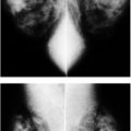
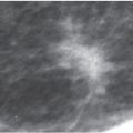
In the 1960s and 1970s, Wolfe was the first to suggest a link between parenchymal patterns on mammography and an increased risk for breast cancer (7–9). Parenchymal pattern descriptors included in the American College of Radiology Breast Imaging Reporting and Data Systems, however, were developed in an attempt to alert clinicians regarding the possible decrease in our ability to detect some cancers in women with “dense” tissue, not as indicators for the risk of breast cancer (14,16,17). The use of these descriptors is subjective with high inter- and intraobserver variability. Additionally, as Kopans (14) points out, it is not possible to accurately measure the percentage of tissue by volume when using two-dimensional mammogram images if exposure values, half-value layers, and compression thickness are not known (e.g., three-dimensional information cannot be obtained accurately using two-dimensional images). In support of this, it is interesting to note that in magnetic resonance imaging (MRI), small amounts of glandular tissue may be seen in some women with seemingly dense tissue mammographically. Additional flaws in these studies include failure to take into account the variability in the positioning of patients for mammograms, the exclusive use of the craniocaudal (CC) projection in some studies, and difficulties in defining the exact extent of the breast and breast tissue (14). It is likely that there is some relationship between the risk for breast cancer and the “density” of the parenchyma on mammography; however, additional studies are needed to further elucidate the exact nature and significance of the relationship.
At this time, other factors reportedly associated with increased breast cancer risk are controversial. These include oral contraceptive use, alcohol intake, exogenous estrogen use (after 10 years of use), breast-feeding, physical activity, and dietary fat intake (saturated fats). In fact, lactation, exercise, and monounsaturated fat intake may have protective benefits. Long-term heavy smoking may be a risk factor, particularly in women who started smoking at an early age (2). Even more controversial risk factors include the use of antiperspirants, bras, induced abortions, and breast implants.
Chemoprevention of breast cancer is controversial and receiving much attention. It may be that drugs such as tamoxifen and raloxifene (selective estrogen receptor modulators [SERMs]) as well as aromatase inhibitors can reduce the risk of developing breast cancer in patients at increased risk. Tamoxifen is approved for use as a breast cancer preventive agent in pre- and postmenopausal women with a significantly increased risk of breast cancer (e.g., patients with atypical ductal hyperplasia or lobular carcinoma in situ). Raloxifene is as effective as tamoxifen in reducing the risk of invasive breast cancer and when compared with tamoxifen, is associated with lower risks of thromboembolic events and cataracts. Tamoxifen is also associated with an increased risk of uterine cancer in women over the age of 50.
SCREENING MAMMOGRAPHY AND SCREENING RECOMMENDATIONS
Mammography is the most studied screening test in medicine, and although there is an abundance of data from randomized controlled trials (RCTs) and service screening studies proving the benefit of screening mammography, the annual use of mammography in women starting at age 40 remains controversial in the minds of some (18,19). The goal of screening mammography is to identify breast cancer as early as possible before it becomes apparent clinically as a lump or with skin changes or distant metastases. But how do we know that mammography can show early breast cancers consistently? And if it can, how do we know that finding these early breast cancers is of any benefit to the patient? The ability of mammography to demonstrate small breast cancers consistently and the benefits of detecting these breast cancers through mammography were established through several RCTs.
Specifically, seven RCTs of screening mammography all of which included women in their 40s have shown a benefit (20–27); two of these were done in North America, one in Scotland, and four in Sweden. Fewer women died of breast cancer among the population invited to screening mammography, compared with the control group of women not invited to undergo screening mammography. The reported benefit ranges from a 20% to 40% reduction in breast cancer mortality among the women invited to screening mammography. Breast cancers are consistently identified through the routine use of screening mammography (20–27). The diagnosis and treatment of women with breast cancers identified through mammography saves lives.
The issue of screening women 40 to 49 years of age has been particularly controversial (28). However, a statistically significant benefit has been reported from several of the RCTs, including the Gothenberg and Malmo trials that reported 44% and 36% reductions in breast cancer mortality, respectively. Also, a statistically significant 26% benefit to screening 40- to 49-year-old women is reported from a meta-analysis (a statistical test that combines data reported from multiple trials) that used data from the seven population-based screening trials (27). In determining appropriate screening intervals, it is important to consider tumor sojourn times. Tumor sojourn time, defined as the time taken for cancers to go from mammographic-preclinical to clinical detectability, is 1.7 years in premenopausal women and 3.3 years in postmenopausal women (29). Screening women in their 40s, therefore, is optimally done at annual intervals.
The Canadian National Breast Screening Study-1 (CNBSS-1) was purportedly aimed at evaluating women in the 40 to 49 age range; however, the flaws in the design and execution of this study are such that the data should not be considered in the same light as those of other RCTs (5,30,31). The two most significant flaws in this study include: (i) The lack of power to show anything less than a 40% decrease in mortality; something that is further impacted by noncompliance (e.g., women in the study group not having screening mammograms) and high contamination (e.g., women assigned to the control group availing themselves of the potential benefits of screening mammography) and (ii) Randomization methodology. The blind randomization of patients into study and control groups is a fundamental requirement of RCTs. For reasons that are unclear, this trial was designed such that a clinical breast examination was done before the randomization of patients into study and control groups. The fact that significantly more women with four or more positive nodes were assigned to the study (mammography) group indicates a serious problem with randomization (32). Additionally, if you consider the over-90% 5-year survival reported for women in the control group, it in essence requires that almost no deaths occur in the study group for a benefit to be shown. As a testament to the fatal randomization issues and resulting imbalances in this trial, the reported 90% 5-year survival in the control group needs to be compared with the 75% 5-year survival in 40- to 49-year-old women in Canada at the time of the study (5). The poor quality of the mammography used for this trial should also be considered. One of the study’s reference physicists stated that the quality of the mammography was not state of the art and that it was even below the quality of mammography being practiced in Canada at that time (33).
Using the cumulative data from all of the randomized controlled screening trials, the American Cancer Society (ACS) in March of 1997 (34) issued new screening guidelines recommending annual screening mammography for all women starting at age 40. This was followed by an update in 2003 (35) that recommends mammography starting at age 40, and advocates that women should be told about the benefits and limitations of breast self-examination (BSE), but states that it is acceptable for women to choose not to do BSE or to do BSE irregularly. The ACS recommends an annual physical examination by a health care provider every 3 years for 20- to 33-year-old women and annually starting at age 40. Currently, the ACS also recommends annual screening MRI for women at high risk, including (36): women with a BRCA mutation and their untested first-degree relatives, women who have had chest wall radiation between the ages of 10 and 30, women with syndromes (Li-Fraumeni, Cowden, and Bannayan–Riley–Ruvalcaba syndromes) associated with an increased breast cancer risk, and those women with a lifetime risk of greater than 20% to 25% (see Fig. 5.36) as determined by risk models (BRCAPRO, Tyrer-Cuzick) (36).
Although there have been no rigorous trials to evaluate the use and benefit of screening mammography in high-risk women, many radiologists recommend starting screening mammography at age 30 for women in a high-risk category including those with a family history of breast cancer (first-degree relative), particularly if the family member developed premenopausal, bilateral, or multiple breast cancers. In a woman whose mother developed breast cancer before the age of 40, mammographic screening should be started 10 years before the age of detection in the mother. So if the mother developed her breast cancer at age 36, mammographic screening in the daughter should be started at age 26.
In 2009, the US Preventive Services Task Force (USPSTF) issued new guidelines (37) for screening mammography every 2 years in 50- to 74-year-old women; they specifically recommended not screening 40- to 49-year-old women or those over the age of 74, and abandoned recommendations for breast self-examination and clinical breast examination (37). The impetus for these recommendations remains unclear since only the Age Trial in England (18), also flawed in design, had been reported since the previous USPSTF guidelines in 2002 (38). Additionally, that computer modeling with rather subjective assumptions would be used to replace the results of RCTs is absurd. The widespread application of these recommendations would be a travesty and likely to reverse the decreases in mortality from breast cancer that have been reported with only 50% of eligible women routinely availing themselves of screening mammography (39). Not only should we be screening women starting at age 40 annually, we should redouble our efforts to encourage more women to avail themselves of this potentially life-saving study starting at age 40; our goal should be 100% compliance with screening recommendations. If there are legitimate issues regarding patient anxiety and false-positive studies, these can be effectively tackled with models such as the ones presented in this book: intense training of radiologists can effectively reduce callback rates to well under 10% and “false-positive biopsies” such that positive predictive values for biopsy recommendations can approximate 50% consistently. Indicated biopsies can be done the day of the recommendation, and if collaborative relationships are established with pathologists and surgeons, pathology results can be obtained within 24 hours of a core biopsy and consultation with a surgeon for definitive treatment can be accomplished within 48 hours of a breast cancer diagnosis. Sadly, it seems to me that the USPSTF is all too willing to throw the baby out with the bathwater in an effort to minimize cost. This is shortsighted and likely to result in the opposite effect. Why are we walking away from the data generated by what in medicine has been the gold standard for establishing proof: the RCTs? RCTs have proven that mammography works starting at age 40, and in the trials as well as in practice, we have seen that its widespread implementation leads to significant decreases in mortality from breast cancer.
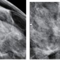
Mammography provides the ability to diagnose stage I or II breast cancers and in many patients when it is at the noninvasive, intraductal stage (stage 0) (40,41). The diagnosis of early, potentially curable breast cancer increases available treatment options for patients and probably renders treatment more effective. Regardless of the histological grade (aggressiveness) of a tumor, or the status of the axillary lymph nodes, women with breast cancers that are less than 1 cm in size, have a 12-year survival rate of 95% (20). The goal of screening mammography, therefore, becomes the identification of cancers that are less than 1 cm in size. Inclusion of all breast tissue in the images through optimal mammographic positioning, high contrast, high resolution and well-exposed images, and the interpretive skills of the radiologist, becomes pivotal in our ability to identify cancers that are less than 1 cm in size consistently.
Mammography is not a perfect test. In some women with breast cancer, the cancer is not visible on the mammogram, even under the best of circumstances. The false-negative rates for mammography range from 7% to 15% (40–42). The sensitivity of mammography is known to be decreased particularly in women with dense tissue mammographically (43,44). This is why it is important that women recognize the need for breast self-examination and annual breast examinations by a health care provider. Although there are false-negative mammograms, screening mammography is an effective method for finding early, potentially curable breast cancers and saving lives. It is the best, and currently the only, reliable defense women have against breast cancer.
MAMMOGRAPHY QUALITY STANDARDS ACT
In 1992, following reports describing a wide range of problems with mammography in the United States, Congress passed the Mammography Quality Standards Act (MQSA) to establish quality standards for mammography. The legislation, authorized for 5 years, was signed on December 12, 1993, and reauthorized on October 8, 1998, extending it into 2002. MQSA regulates mammographic modalities defined as technologies for radiography of the breast. Stereotactic biopsies, needle localizations, and ductography are procedures currently exempt from the definition of mammographic modality and not regulated under MQSA; ultrasound, MRI, and nuclear medicine studies are non-radiographic procedures and not regulated under MQSA.
Effective October 1, 1994, all mammography facilities (except those of the Department of Veterans Affairs) must: (i) Get accredited by an approved accrediting body every 3 years; (ii) Get certified by the Secretary of Health and Human Services (HHS) every 3 years; (iii) Undergo annual on-site inspection by Food and Drug Administration (FDA)–trained and certified federal or state inspectors on behalf of HHS.
The current FDA-approved accrediting bodies include the American College of Radiology (ACR) and the States of Arkansas, Iowa, and Texas. The last update of MQSA statistics on the FDA website (www.fda.gov) reports 8,698 certified facilities as of November 1, 2013, with 13,053 accredited units; 7,970 facilities are certified with full field digital mammography (FFDM) units representing 12,100 accredited FFDM units. As of the November 1, 2013, update available on the FDA website, 88% of inspected facilities were in compliance; 0.2% of inspected facilities had level I noncompliance violations requiring immediate action for remedy, reinspection, and sanctions if corrective actions are not taken; 10.8% had level II violations requiring a written response with corrective actions required within 30 days, and 1% had level III violations requiring corrective action before the next inspection. As of October 1, 2007, the inspection fee for facilities is $2,150 for the first unit and $250 for each additional unit. The fee for facilities requiring reinspection is $1,140.
Final MQSA regulations are in effect as of April 28, 1999. The Federal Register publication of October 28, 1997 (45) provides the final regulations. This law regulates mammographic facilities, including initial and continuing qualification standards for interpreting physicians, radiologic technologists, medical physicists, and mammography facility inspectors. The law spells out quality standards for mammographic equipment and practices, what is needed for the quality assurance (QA)/quality control (QC) program, record keeping, mammography reports, as well as patient and referring physician notification. Familiarity with the details is recommended, particularly for those designated as the lead interpreting physician (please refer to the Federal Register publication for all the nuances of the law—beyond the scope of this book).
AMERICAN COLLEGE OF RADIOLOGY MAMMOGRAPHY ACCREDITATION PROGRAM
Since the late 1980s, the ACR has taken a leadership role in addressing mammographic image quality. The Mammography Accreditation Program (MAP) developed by the ACR is now the oldest and largest accreditation program in the United States. The MAP program accredits units (FDA certifies facilities) every 3 years. For accreditation facilities, submit: (i) Completed form providing information on equipment, personnel qualifications, and test image data; (ii) Phantom image for image quality and dose evaluations, using an approved phantom and thermoluminescent dosimeter; (iii) Two normal mammograms (fatty and dense breasts); (iv) Processor quality control for a 30-day period; and (v) Fee (initial cycle and renewal $1,475 for first unit and $1,300 for each additional unit) (46).
Facilities are also required to provide the ACR with an annual update. This update includes QC documentation, the medical physicist’s annual survey for each unit, and an application data update (e.g., changes in personnel). Under MQSA, the ACR is required to perform on-site surveys of at least 5% of the facilities it accredits; 50% of these are selected randomly and the others based on problems identified through FDA inspections, serious consumer complaints, or history of noncompliance. A medical physicist, radiologist, and an ACR staff person make up the survey team. Additionally, at least 3% of accredited facilities are randomly selected to undergo a random clinical image review (46).
The ACR has subsequently developed accreditation programs for stereotactic breast biopsy, diagnostic breast ultrasound, ultrasound-guided breast biopsy, and breast MRI. The ACR now also provides a “Breast Imaging Center of Excellence designation” to those facilities that are ACR-accredited in mammography, stereotactic breast biopsies, breast ultrasound, ultrasound-guided biopsy, and breast MRI. The readers are strongly encouraged to familiarize themselves with these programs by contacting the ACR or visiting their website (www.ACR.org).
TREATMENT OPTIONS
The treatment of breast cancer usually entails a combination of surgery, radiation therapy, and chemotherapy. Surgical options include mastectomy or lumpectomy. With the exception of some patients with localized DCIS, sampling of the axillary lymph nodes with a sentinel lymph node (SLN) biopsy or axillary lymph node dissection (ALND) is done at the time of the patient’s definitive surgery. Immediate or delayed reconstruction with an implant or autologous tissue transplantation is always an option that should be discussed with patients undergoing mastectomy; in these patients, reduction of the contralateral breast may also be appropriate.
Needless to say, mastectomy is always an option for patients; however, depending on the size of the lesion relative to the size of the breast, lumpectomy and radiation therapy (see below) are as effective as mastectomy in treating most patients with localized breast cancer. The goal of lumpectomy is to remove the entire tumor so there is little, if any, cancer left in the breast with, ideally, a cosmetic result that is acceptable to the patient. The margins of the lumpectomy specimen are evaluated histologically, and if disease extends to the margins, re-excision of the tumor bed is usually done as a separate surgical procedure. The goal of re-excision is to get negative margins. If the margins are grossly positive after re-excision, the patient may not be a good candidate for conservative therapy and mastectomy may be more appropriate. The increasing use of breast MRI to delineate the extent of disease at the time of diagnosis may facilitate preoperative surgical decisions since it can help distinguish patients with extensive or multifocal disease needing mastectomy from those with localized lesions who are more amenable to lumpectomy.
Historically, patients with invasive breast cancer had full ALNDs involving the excision of multiple (but a variable number) lymph nodes from the axilla. Complications associated with ALND include lymphedema, arm and shoulder numbness, paresthesias involving the arm and shoulder, loss of range of motion at the shoulder, and the formation of fluid pockets (seromas, hematomas) in the axilla. Many surgeons have adopted SLN biopsies to initially stage patients with clinically node-negative breast cancer. The limited dissections needed for SLN biopsies have minimized the complications associated with ALND and, as such, have made a significant contribution to patient care and overall quality of life for patients.
The concept of the SLN biopsy is fairly simple and first developed in the evaluation of patients with melanoma. If the lymphatic drainage from the breast to the axilla proceeds in an orderly, stepwise manner, one or two lymph nodes in the axilla should receive the lymphatic drainage from the breast first and consistently. If these lymph nodes can be identified reliably, and if they reflect the overall status of the axillary lymph nodes accurately, axillary dissections can be limited to the removal of the SLNs. To identify the SLN, a vital blue dye, technetium-labeled sulfur colloid particles, or both are injected subcutaneously, peritumoral or peri-areolar. Visual inspection of the axilla by the surgeon for collection of vital blue dye, or radioactivity detected with a handheld gamma probe, is used to identify one, two, and sometimes three nodes considered the SLN(s) (47–50). The use of both methods increases the likelihood of identifying the SLN in up to 90% of patients with an overall accuracy of 98.2% and a false-negative rate of 5.8% (51). In those patients in whom the SLNs cannot be mapped an ALND is done.
The use of SLN biopsy is indicated in patients with T1- and T2-sized tumors, multicentric disease, DCIS when mastectomy is planned, in older and obese patients and in men with breast cancer. Although not always done, efforts can be made to assess (imprint or touch prep, cytology of cells scraped from the cut surface of node, or frozen section) the sampled lymph node at the time of surgery: if metastatic disease is identified with the patient still in the operating room, the surgeon can do the ALND. If the touch prep is not done at the time of surgery, or if it is interpreted as negative, and metastatic disease is identified on the permanent hematoxylin and eosin (H & E) stained slides of the SLN, the patient is scheduled for a completion ALND since approximately 43% of these patients are found to have additional node disease on the ALND (52).
The significance of isolated tumor cells (ITC) and micrometastases (>0.2 mm but not >2 mm) in axillary lymph nodes remains unclear. Reportedly, metastases are identified in non-sentinel lymph nodes in 10% of patients with ITCs and in 20% to 35% of patients with micrometastatic disease in the SLN (52). Currently, the American Society of Clinical Oncology (ASCO) recommends ALND in patients with micrometastases in the SLN regardless of the method of detection (52).
Patients with large or locally advanced invasive breast cancers, inflammatory carcinoma, or in whom axillary disease is suspected clinically, are not considered good candidates for SLN biopsy and, as such, undergo ALND. In pregnant patients, SLN biopsies are done using the technetium labeled sulfur colloid but not the blue dye. In patients with prior breast surgery and invasive disease, SLN biopsy is attempted, however, if the nodes do not map an ALND is indicated. Several major questions remain unanswered with respect to sentinel node biopsies in women with breast cancer. These include: (i) Which patients with positive SLN biopsies can be treated with breast or axillary radiation, and which patients need completion ALND; (ii) The clinical significance of negative SLN biopsy after neoadjuvant therapy; and (iii) What is the clinical significance of ITCs, and do these patients need an ALND? (52).
Radiation therapy for local control is usually recommended following lumpectomy. In a small number of patients, radiation therapy to the chest wall or axilla is also recommended following mastectomy to reduce the likelihood of chest wall or axillary recurrence. Whole breast irradiation is usually started 2 to 5 weeks after the lumpectomy and is given 5 days a week for 6 weeks (40 to 50 Gy). On the basis of the size of the tumor, patient age, disease-free surgical margins, and in those with negative axillary lymph nodes, accelerated partial breast irradiation (APBI) may be recommended in some patients. This entails high doses of radiation localized to the lumpectomy site commonly given over a 5-day period. APBI methods under investigation include: (i) Intracavitary brachytherapy using an inflatable balloon catheter placed in the lumpectomy bed intraoperatively or percutaneously after the lumpectomy; (ii) Interstitial brachytherapy using multiple afterloading catheters placed around the lumpectomy bed and (iii) Intraoperative radiation therapy.
In thinking about chemotherapy consider three major groups of patients: (i) Those with hormone receptor–positive cancers who can be managed with receptor targeted therapy ± chemotherapy (see discussion on Oncotype); (ii) Those with HER2/neu positive tumors in whom trastuzumab (and Lapatinib approved for patients with HER2/neu positive metastatic disease) can be used in conjunction with chemotherapy; and (iii) Those with estrogen (ER) negative, progesterone (PR) negative, HER2/neu negative tumors (triple negative tumors; TNT) in whom chemotherapy is the only systemic treatment available.
Chemotherapy is usually recommended if there is metastatic disease to the axillary lymph nodes or in patients with distant metastases. In patients with node-negative estrogen receptor–positive tumors, the use of the Oncotype DX assay reportedly provides prognostic information with respect to the likelihood of recurrence and response to chemotherapy (CMF = cyclophosphamide, methotrexate, and fluorouracil) (53). Oncotype DX is a reverse transcriptase/polymerase chain reaction assay based on the analysis of a 21-gene panel. It is used to calculate a recurrence score (RS) of 0 to 100. Patients with a low RS (RS < 18) have indolent tumors that are sensitive to hormonal therapy, and chemotherapy adds little, if any, benefit (53). Patients with a high RS (RS ≥ 31) have aggressive tumors less likely to respond to hormone therapy, and benefit significantly from adjuvant therapy with a decrease in the recurrence rate at 10 years of 27.6% (53). In patients with an intermediate recurrence score (RS ≥18 to 30), it is unclear whether the benefits of chemotherapy exceed the risks (53).
Neoadjuvant therapy is recommended in patients with larger sized tumors, locally advanced breast cancer, or inflammatory carcinoma and is being increasingly used in women with larger lesions in whom lumpectomy may become an option after therapy. Depending on the response of the tumor, neoadjuvant therapy can be followed by mastectomy or, in selected patients, lumpectomy. Radiation therapy and additional chemotherapy may be recommended in these patients after their definitive surgery.
The 5-year relative survival rate for women diagnosed with lymph node–negative invasive lesions is 98%; survival rates decrease to 84% and 24% in patients with metastatic disease to regional lymph nodes and those with distant metastatic disease, respectively. For all breast cancer stages combined, relative survival rates at 10 and 15 years are 83% and 77%, respectively. Five-year relative survival rates for invasive lesions have improved from 75% in the mid-70s to 90% in 2014 (2).
BREAST CANCER STAGING
Women diagnosed with breast cancer are staged based on the size of the tumor (T), the presence or absence of regional lymph node (N) involvement, and the presence or absence of cancer cells at sites distant (M) to the breast. This system is referred to as the TNM system (54). Numbers are assigned to each of these letters to define the characteristics of the cancer found in an individual patient.
Clinical staging (cTNM) is based on information obtained from the clinical evaluation of patients, including a complete physical examination, imaging (except lymphoscintigraphy), biopsy, and surgical exploration before any treatment is given (see Tables 1.1 and 1.2). Pathological staging (pTNM) is based on information from the clinical evaluation and complete removal and evaluation of the primary tumor and regional lymph nodes (see Table 1.3) following surgery but before any treatment is given (20). After each one of these three components (TNM) is classified (see Table 1.4 for classification of “M”), the patient is assigned into one of several stages (see Table 1.5). Recommendations for treatment, and the overall prognosis of a patient, vary depending on the stage of the breast cancer at the time of diagnosis. Post-neoadjuvant therapy TNM findings are designated by the prefix “yc” if clinical (ycTNM) or “yp” if pathological (ypTNM). Currently, no stage is provided for patients who have a complete pathological response following neoadjuvant therapy (ypT0ypN0cM0).
Axillary, internal mammary, and supraclavicular lymph nodes on the side (ipsilateral) of the primary tumor are considered regional lymph nodes in patients with breast cancer. Level I (low axilla) axillary lymph nodes are defined as those lateral to the lateral margin of the pectoralis minor muscle. Level II (mid axilla) axillary lymph nodes are those found between the medial and lateral borders of the pectoralis minor muscle; this includes interpectoral nodes (e.g., Rotter lymph nodes). Level III (apical axilla) axillary lymph nodes are those found medial to the medial margin of the pectoralis minor muscle and inferior to the clavicle (aka apical or infraclavicular nodes). For the purposes of classification and staging, intramammary lymph nodes are considered axillary lymph nodes. If metastatic disease is identified in cervical, contralateral axillary, or internal mammary lymph nodes, it is considered distant disease (M1).
Table 1.1 PRIMARY TUMOR (T) CLASSIFICATION
Related posts:

Stay updated, free articles. Join our Telegram channel

Full access? Get Clinical Tree


