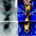
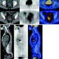
Fig. 44.1
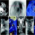
Left subclavear adenopathy measuring less than a centimeter
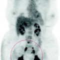
In the left para-aortic and celiac-mesenteric areas and hepatic hilum, there are numerous confluent enlarged lymph nodes, characterized by high glucose metabolism, SUVmax 8.5.
The pulmonary nodule shown in the anterior segment of the left lower lobe and previously radio-treated, shows limited consumption of glucose due to actinic damage, SUVmax 1.8 (Fig. 44.1, green arrow).
Mild metabolic alteration to the thoracic spine (D6 and D10) due to benign disease, SUVmax 2.2.
44.4 Conclusions
The PET scan shows progression of disease due to extended lymph node involvement.
There were no focal lesions of the liver and lung with high metabolism. See Figs. 44.2, 44.3, 44.4, 44.5, 44.6.

Fig. 44.2


CT scan shows, in the anterior segment of the lower lobe of the left lung, a metastatic mantle nodule, which had been previously radio treated. The PET scan showed restricted carbohydrate consumption due to late post-actinic remodeling
Stay updated, free articles. Join our Telegram channel

Full access? Get Clinical Tree


