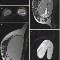
Fig. 1.1
Sagittal T1 fat-saturated pre-contrast image of the right breast

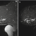
Fig. 1.2
Sagittal T1 fat-saturated post-contrast image of the right breast (a) and corresponding T1 non–fat-saturated image (b)

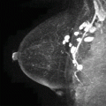
Fig. 1.3
Sagittal T1 non–fat-saturated image of the left breast

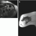
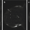
Fig. 1.4
Sagittal T1 non–fat-saturated image of the right breast (a) and corresponding T1 fat-saturated sequences (b)
1.2 T1-Weighted Sequence
Teaching Points
Breast MRI should be performed on systems with at least 1.5 Tesla magnet strength with a dedicated breast coil. Usually, a pre-contrast T1-weighted gradient-echo sequence without fat saturation is the first sequence performed after the scout images. A T1-weighted non–fat-saturated sequence is obtained bilaterally, including axillae and chest wall, to distinguish fat from water-based tissues. The pre-contrast T1-weighted sequence is typically used to assess the amount of fibroglandular tissue and can be particularly helpful in documenting the presence of high-signal hemorrhagic or proteinaceous fluid-filled, dilated ducts (Fig. 1.1). Radiologists should be aware of these T1-hyperintense ducts, which can cause potential misregistration artifact related to patient motion, leading to an apparent suspicious linear nonmass enhancement on post-contrast subtracted images. It is important to assess the pre-contrast T1 sequence before evaluating the post-contrast images. T1 non–fat-saturated images can be helpful in evaluating for fat necrosis by identifying central fat (Fig. 1.2). Biopsy clip artifact and architectural distortion can be better seen on the non–fat-saturated T1 sequence (Figs. 1.3 and 1.4).
Image Findings

Fig. 1.5
Hemorrhagic or proteinaceous debris in ducts. Sagittal T1 fat-saturated pre-contrast image of the right breast demonstrates several linear hyperintensities (arrows) in a ductal distribution in the lower breast, compatible with hemorrhagic or proteinaceous debris

Fig. 1.6
Fat necrosis. (a) Sagittal T1 fat-saturated post-contrast image of the right breast demonstrates indeterminate rim enhancement (arrows). (b) The T1 non–fat-saturated image shows central fat (arrow), compatible with benign fat necrosis

Fig. 1.7
Architectural distortion. In a patient with prior lumpectomy, the sagittal T1 non–fat-saturated image of the left breast illustrates architectural distortion in the central breast (arrows) and associated nipple retraction (arrowhead)

Fig. 1.8
Susceptibility artifact from biopsy clip. The susceptibility artifact from a biopsy clip (arrow) is more prominent on the T1 non–fat-saturated image of the right breast (a) than on the T1 fat-saturated sequences (b)
1.3 History
Four patients illustrating the range of the amount of fibroglandular tissue on breast MRI (Figs. 1.9, 1.10, 1.11, and 1.12).

Fig. 1.9, 1.10, 1.11, and 1.12
Selected sagittal T1 post-contrast image (a) right breast demonstrates an almost entirely fatty breast (b) right breast shows scattered fibroglandular tissue. Selected sagittal T1 pre-contrast image (c) right breast demonstrates heterogeneous fibroglandular tissue. Selected sagittal T1 post-contrast image (d) left breast demonstrates extreme fibroglandular tissue
1.4 Fibroglandular Tissue
Teaching Points
At mammography, breast density is represented by the amount of fibroglandular tissue (FGT) in contrast to fat measured in two-dimensional views that may not reflect an accurate assessment. Breast MRI provides strong soft-tissue contrast between FGT and fat, and a more accurate three-dimensional coverage of the entire breast. Evaluation of the amount of FGT is usually done on the T1-weighted sequences with and without fat suppression. According to the American College of Radiology (ACR) guidelines, the amount of FGT should be described in the Breast MRI report using the BI-RADS® four assessment categories of breast composition, which are defined by the visually estimated content of FGT within the breasts.
The amount of fibroglandular tissue (breast density) has been established as an independent risk factor associated with the development of breast cancer, which is three to five times higher in women with mammographically dense breasts than in women with predominantly fatty breasts.
1.5 History
Four patients illustrating the range of background parenchymal enhancement seen on breast MRI (Figs. 1.13, 1.14, 1.15, and 1.16).

Fig. 1.13,1.14, 1.15, and 1.16
Sagittal T1 post-contrast 3D maximum intensity projection (MIP) image (a) left breast demonstrates minimal background parenchymal enhancement (b) right breast demonstrates mild background parenchymal enhancement (c) right breast demonstrates moderate background parenchymal enhancement (d) right breast demonstrates marked background parenchymal enhancement
1.6 Background Parenchymal Enhancement
Teaching Points
The 5th edition of the ACR BI-RADS® (Breast Imaging Reporting and Data System) Atlas recommends that the amount of background parenchymal enhancement (BPE) be categorized in the MRI report as “minimal,” “mild,” “moderate,” or “marked” to describe the volume and intensity of the BPE. Research into the association of BPE and breast cancer risk indicates a likely increase in the risk for breast cancer, which may be as high (3–4×) as the more-established risk associated with mammographic dense breasts.
1.7 History
67-year-old woman with newly diagnosed right breast cancer (Figs. 1.17, 1.18, 1.19, and 1.20).


Fig. 1.17
T1-weighted fat-saturated pre-contrast image (a) and three consecutive post-contrast images (b–d) demonstrate 1.5-cm, mildly irregular enhancing mass in the anterior upper right breast
Fig. 1.18
Close-up evaluation of the mass (a) demonstrates the highest signal intensity of the mass on the first post-contrast image (b), with subsequent decrease in signal intensity on the delayed images (c, d). (e) Computer-generated curve of the signal intensity time course of the mass. (f) The pattern of washout is color-coded red
1.8 Computer-Aided Detection
Teaching Points
In addition to the morphology assessment, the enhancement kinetics pattern can be analyzed by obtaining at least three post-contrast images. Typical breast tumor demonstrates intense and rapid uptake of contrast owing to increased angiogenesis and washout of contrast owing to increased capillary permeability and shunting. A study by Kuhl et al. (2005) showed kinetic analysis, specifically the signal intensity time course, to be specific for malignancy.
Three types of signal intensity time course (Fig. 1.19) can be identified:
Type 1: Progressive
Type 2: Plateau
Type 3: Washout
When mixed enhancement is present, the worst curve is reported (Fig. 1.20).
Of note, the morphology assessment should outweigh the kinetics analysis in clinical decision making. Morphology has higher specificity for malignancy.
Related posts:

Stay updated, free articles. Join our Telegram channel

Full access? Get Clinical Tree


