Bursitis
KEY FACTS
Terminology
Imaging
IMAGING
General Features
 Subacromial-subdeltoid bursa: Large bursa overlying rotator cuff tendons
 Olecranon bursa: Overlies olecranon process & distal end of triceps tendon
 Ischial tuberosity bursa: Between ischial tuberosity (hamstring tendon attachment) & gluteus maximus muscle
 Iliopsoas bursa: Large bursa between iliopsoas tendon & anterior part of hip joint
 Trochanteric bursa: Laterally overlies gluteus medius & minimus tendon insertion into greater trochanter femur
 Subgluteus minimus bursa: Between gluteus minimus tendon & anterior facet greater trochanter
 Subgluteus medius bursa: Between gluteus medius tendon & posterosuperior facet greater trochanter
 Semimembranous bursa: Posteromedial to semimembranous musculotendinous junction & tendon posteromedial aspect distal thigh
 Pes anserine bursa: Between sartorius, gracilis, semitendinosus tendons, & posteromedial tibia
 Prepatellar bursa: Overlies inferior pole of patella & proximal patellar tendon
 Infrapatellar bursa: Overlies mid-to-distal aspect of patellar tendon
 Retrocalcaneal bursa: Between calcaneus, Achilles tendon, & Kager fat pad
 Superficial calcaneal bursitis: Overlies distal aspect of Achilles tendon
Ultrasonographic Findings
 Majority of bursae not visible on US unless distended with fluid or proliferative synovial tissue
 Distended hypoechoic bursa
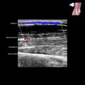
 Acute bursitis: Thin-walled bursa distended with hypoechoic fluid, peribursal hyperemia
 Chronic cases: Greater thickening of bursal wall, more mature synovial proliferation, more echogenic content, & increased likelihood of intrabursal hyperemia


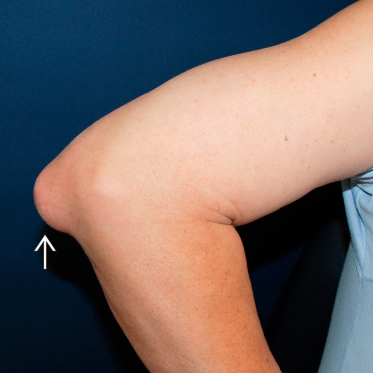
 that has been present for years but recently increased in pain and size.
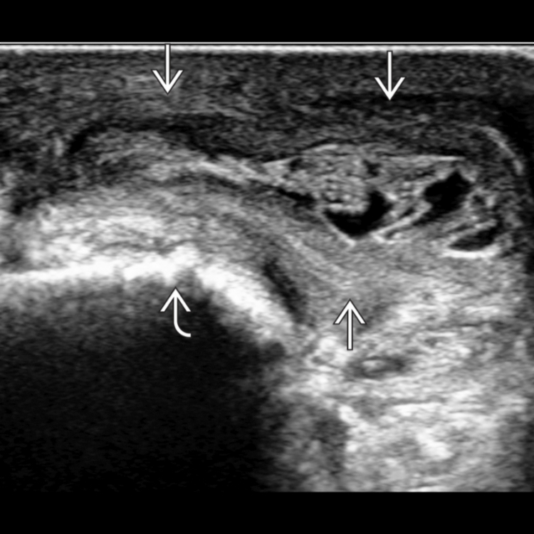
 . The bursa is distended by fluid and synovial proliferation and overlies the olecranon process
  . Olecranon bursae frequently have a thickened wall, internal debris, and septa. A noninflamed olecranon bursa is not visible on US.
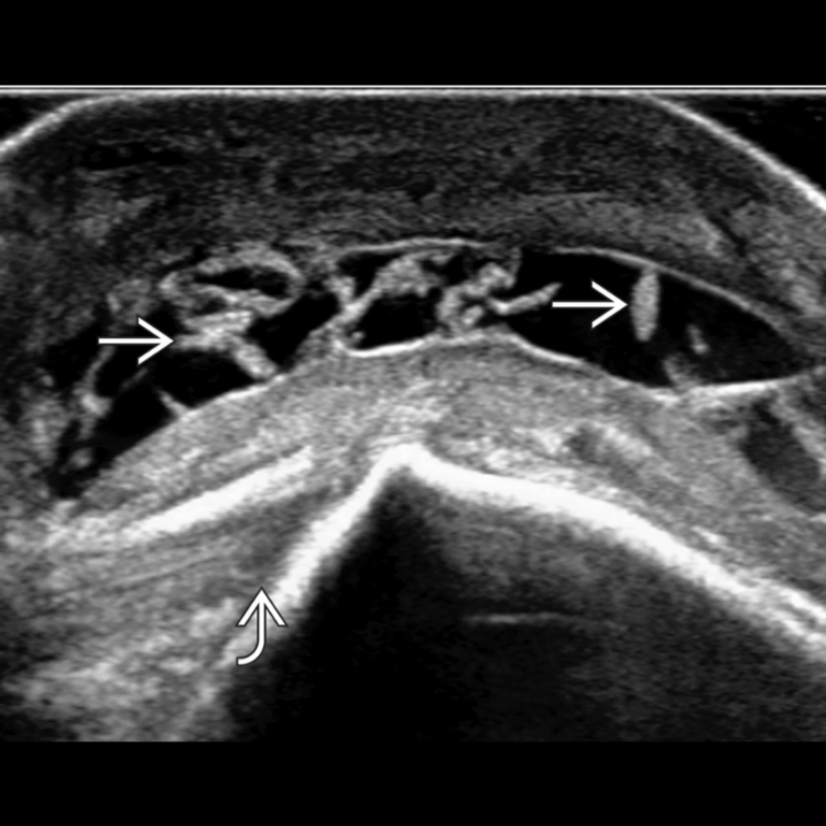
 , giving the bursa a multiloculated appearance. The normal triceps tendon insertion
  into the olecranon process is shown.
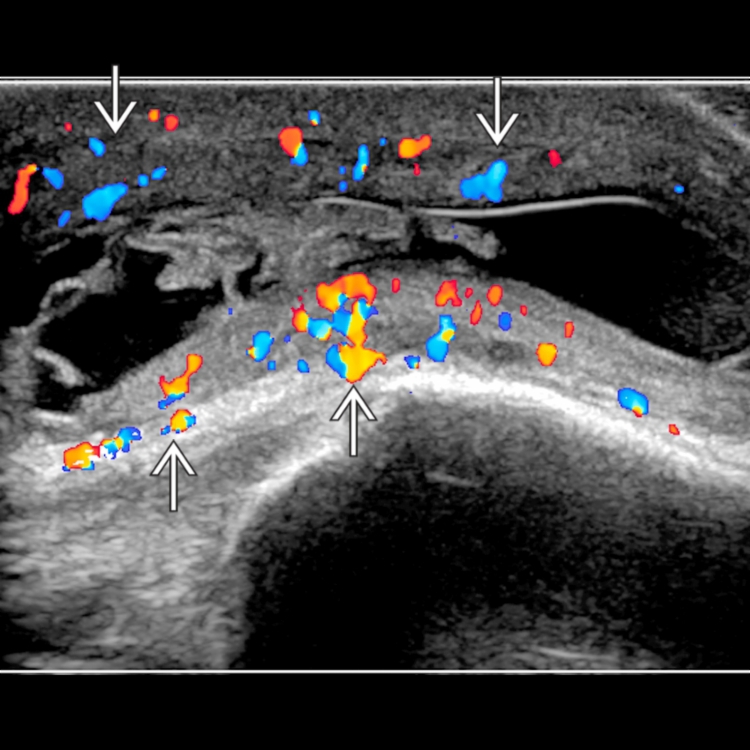
 and peribursal tissues. The internal content and septations show no hyperemia.