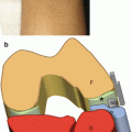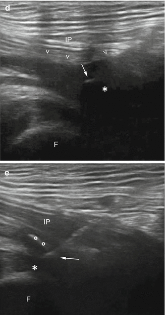
Fig. 6.1
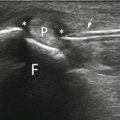
US-guided treatment of iliopsoas bursitis on a long axis. (a) Probe and patient position to perform US-guided treatment of iliopsoas bursitis. (b) Anatomical scheme and (c) US scan of iliopsoas bursitis treatment. F femur, IP iliopsoas muscle, asterisk bursa, arrow needle tip. (d) US image taken during aspiration of the bursa. Arrowheads show the bowing of the superficial surface of the bursa. (e) End of the procedure; the bursa is completely drained and steroid has been injected inside (circles)
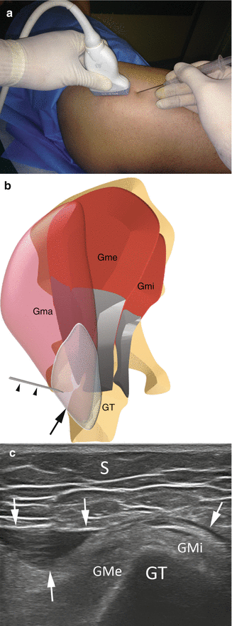
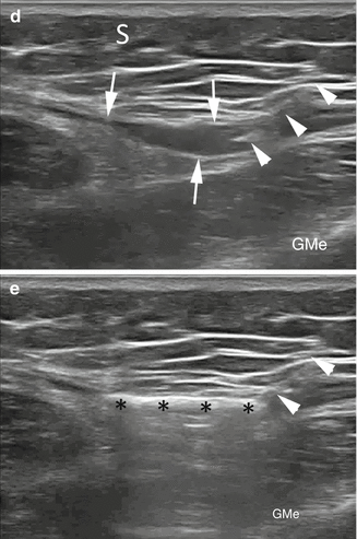
Peritrochanteric bursitis: the patient lies on the contralateral side. The bursa is demonstrated with an axial scan over the greater trochanter avoiding applying excessive pressure. The needle can be inserted with a posterior-to-anterior, medial-to-lateral, in-plane approach. In the case of distention of the gluteus medius or minimus bursae, an anterior-to-posterior insertion of the needle is necessary. The procedure is shown in Fig. 6.2.