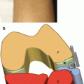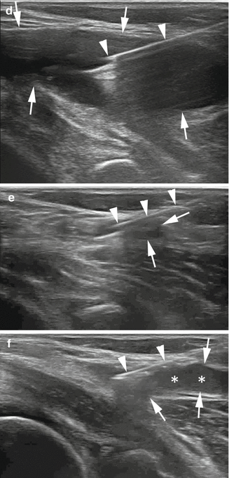
Fig. 9.1
US-guided treatment of gastrocnemius bursitis on a long axis. (a) Probe and patient position to perform US-guided treatment of gastrocnemius bursitis. (b) US image of a large gastrocnemius bursitis (arrows). (c) Anatomical scheme and (d) long-axis US scan of gastrocnemius bursitis. LG lateral gastrocnemius muscle, MG medial gastrocnemius muscle, SM semimembranosus tendon, arrowheads needle. (e) End of the procedure; the bursa is completely drained. (f) Steroid injection (asterisks)
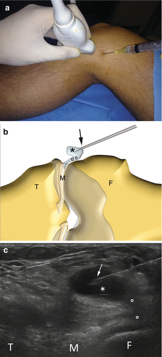
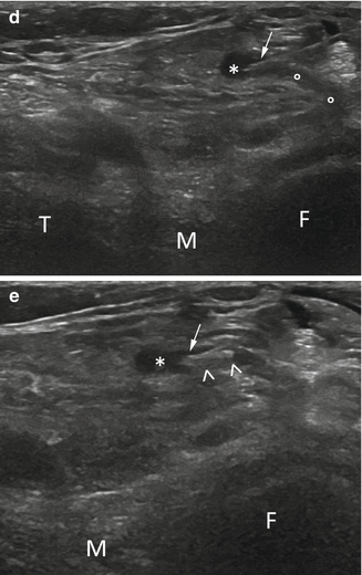
Parameniscal cysts: the patient is positioned according to the location of the cyst. Usually, the needle is inserted according to the major axis of the cyst. The procedure is shown in Fig. 9.2 . When the cyst involves the superficial peroneal nerve, extreme caution should be taken to avoid the nerve fascicles. The procedure is shown in Fig. 9.3 .
