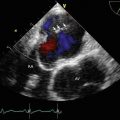
Fig. 3.1
Transthoracic echocardiography (TTE), parasternal long-axis view showing a mass with round, smooth borders and a heterogeneous echo density located at the posterior mitral annulus (arrows). Ao aorta, LA left atrium, LV left ventricle, RV right ventricle
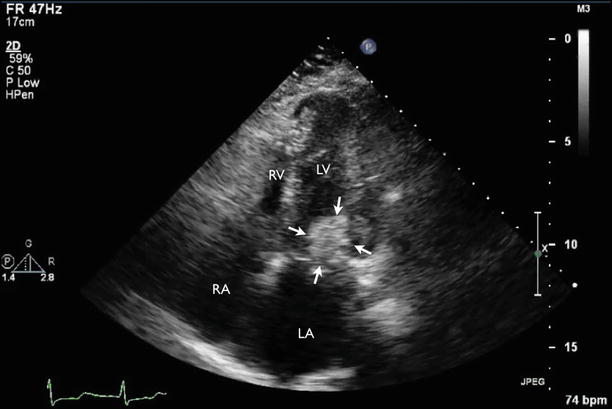
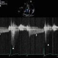
Fig. 3.2
TTE, apical four-chamber view showing a posterior mitral annulus mass (arrows). RA right atrium
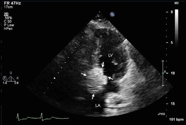
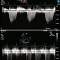
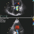
Fig. 3.3
TTE, apical two-chamber view showing a posterior mitral annulus mass (arrows)
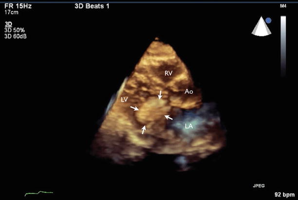
Fig. 3.4
Three-dimensional (3D) TTE, parasternal long-axis view again showing a mass with round, smooth borders and a heterogenous echo density located at the posterior mitral annulus (arrows)
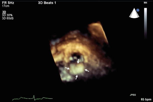
Fig. 3.5
3D TTE, mitral valve viewed from left atrium showing posterior mitral annulus prominence (arrows)
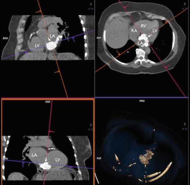
Fig. 3.6
Cardiac CT scans with multiplanar reconstruction confirmed the diagnosis and showed an intracardiac mass measuring 3.4 × 4.7 cm involving both anterior and posterior mitral annulus with extension into the left atrium (LA) wall (arrow). The periphery of this mass is densely calcified in spots, whereas the central portion has density of approximately 400 Hounsfield units, consistent with liquefaction of calcified mitral annulus
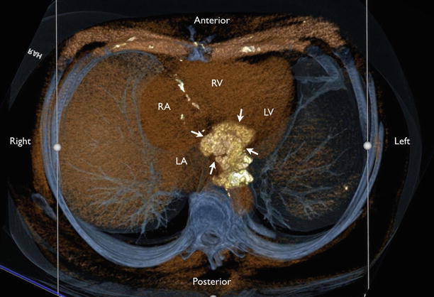
Fig. 3.7
3D CT reconstruction showing the extension of the mitral annular calcification mimicking a cardiac mass (arrows)
Video 3.1 Transthoracic echocardiography (TTE), parasternal long-axis view showing a mass with round, smooth borders and a heterogeneous echo density located at the posterior mitral annulus (AVI 5136 kb)
Video 3.2 TTE, apical four-chamber view showing a posterior mitral annulus mass (AVI 6292 kb)
Video 3.3 TTE, apical two-chamber view showing a posterior mitral annulus mass (AVI 5368 kb)
Video 3.4 Three-dimensional (3D) TTE, parasternal long-axis view again showing a mass with round, smooth borders and a heterogenous echo density located at the posterior mitral annulus (AVI 1511 kb)
3.1.1 Learning Points
Caseous calcification of the mitral annulus (CCMA) is a rare variant of mitral annular calcification with an estimated prevalence of 0.068 % [1–3]. It is characterized by an echodense outer shell and echolucent core [4]. The posterior mitral annulus is more often affected than the anterior. The inner core of the lesion represents the byproduct of liquefaction necrosis. Pathological examination reveals sterile, amorphous, acellular eosinophilic material with macrophage and lymphocyte infiltration [5].
The clinical course of CCMA is generally benign and often dynamic, with reports of spontaneous resolution of progression [3]. The complications of CCMA include hemodynamically significant mitral stenosis and regurgitation secondary to mass effect [5, 6], as well as erosion of the inner core into the left atrial chamber [7] and the left circumflex coronary artery [8].
3.2 Case 2. Calcific Mitral Stenosis
A 55-year-old man with end-stage renal failure secondary to Alport’s disease who is undergoing thrice-weekly hemodialysis reports increasing breathlessness with minimal exertion and palpitations. He was noted to have a murmur on auscultation (Figs. 3.8, 3.9, 3.10, 3.11, 3.12, 3.13, and 3.14).
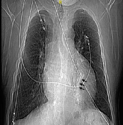
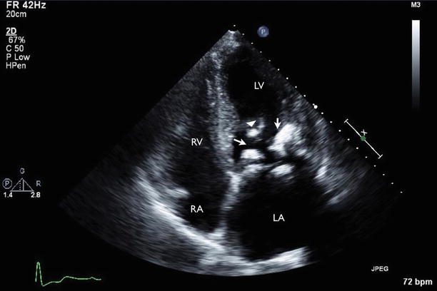
Fig. 3.8
Posteroanterior (PA) chest x-ray showed mitral annular calcification (arrows) and scoliotic curvature of the thoracolumbar spine, convex to the right
Fig. 3.9


TTE, apical four-chamber view showed bright echodensities along the mitral annular plane involving both anterior and posterior aspects of the annulus (arrows), as well as calcified subvalvular apparatus (arrowhead)
Related posts:

Stay updated, free articles. Join our Telegram channel
Full access? Get Clinical Tree
