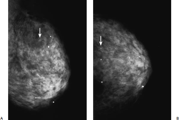
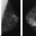
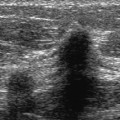
14 Calcifications: Coarse/Popcorn-Fibroadenoma A 69-year-old woman presents for screening mammogram. • Normal exam Calcifications (Figs. 14.1 and 14.2) • Type: coarse/popcorn • Distribution: single Fig. 14.1 Faint coarse calcifications are present in the upper outer breast (arrow). (A) Left MLO mammogram. (B) Left CC mammogram. Fig. 14.2 Exam performed 3 years after Fig. 14.1. The calcifications (arrow) have enlarged and are now characteristic of an involuting fibroadenoma. (A) Left MLO mammogram. (B) Left CC mammogram. • Fibroadenoma • BI-RADS assessment category 2, benign finding • Differential diagnosis of large, coarse calcifications includes fibroadenoma, fat necrosis, and calcium deposition from hypercalcemia (i.e., hyperparathyroidism, renal failure). Gershon-Cohen J, Ingleby H. Roentgenography of fibroad-enoma of the breast. Radiology 1952;59:77–87 Han SY, Witten DM. Diffuse calcification of the breast in chronic renal failure. AJR Am J Roentgenol 1977;129:341–342
Case 14.1: Coarse/Popcorn–Fibroadenoma
Case History
Physical Examination
Mammogram
Pathology
Management
Pearls and Pitfalls
Suggested Reading
Case 14.2: Coarse/Popcorn–Fibroadenoma
Case History

Related posts:


Stay updated, free articles. Join our Telegram channel

Full access? Get Clinical Tree


