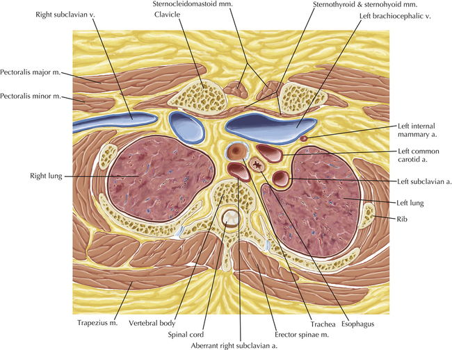
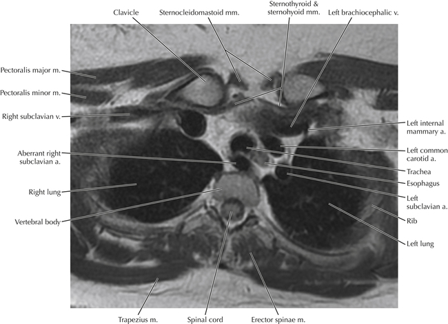
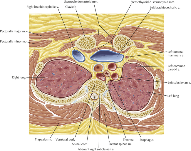
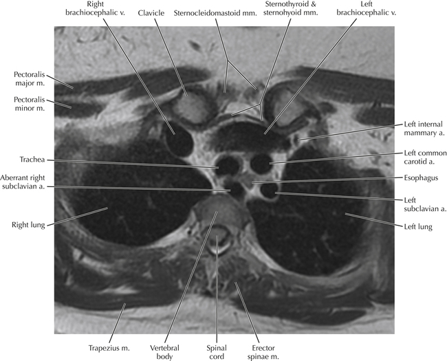
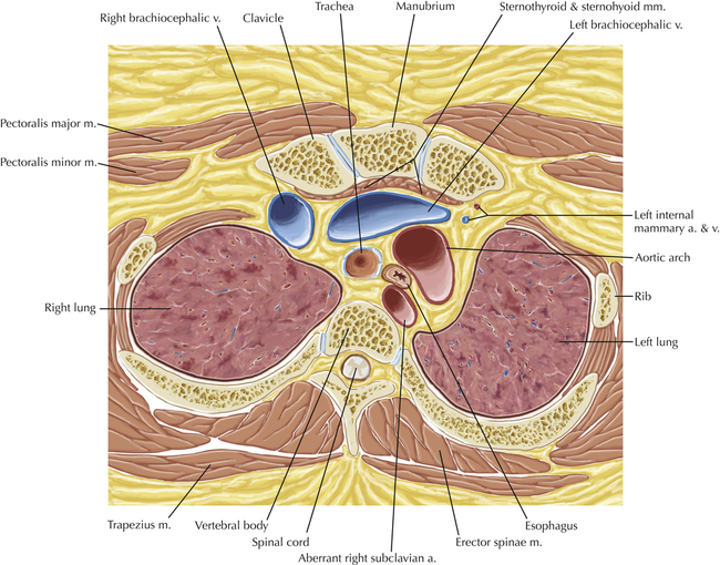
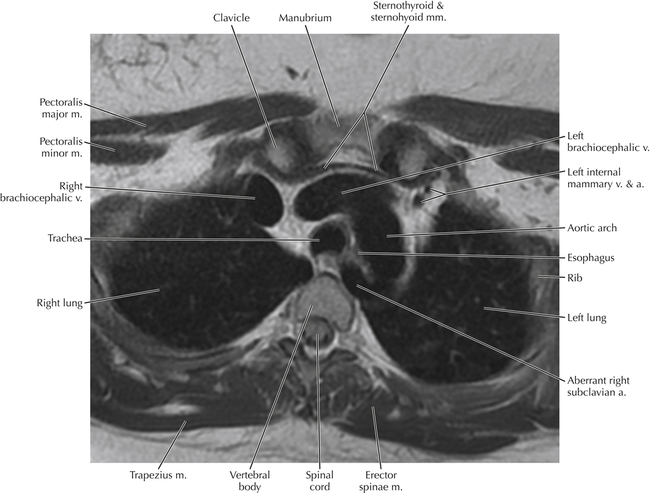
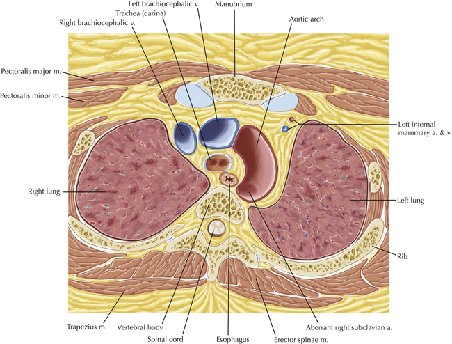
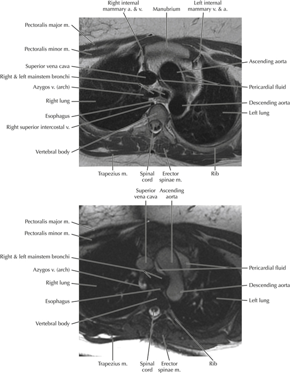
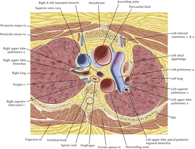
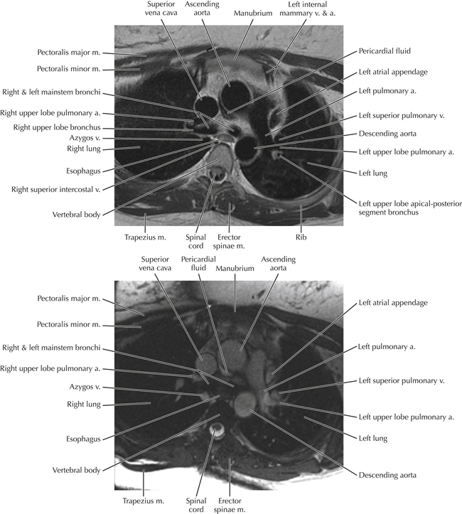
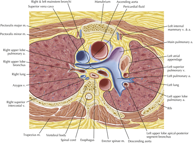
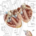
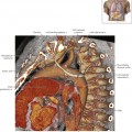
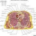
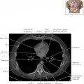
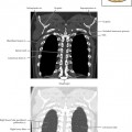
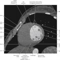
Chapter 9 Cardiac Anatomy Using MR AXIAL 2-CHAMBER 3-CHAMBER 4-CHAMBER SHORT AXIS Cardiac MR Axial 1 Normal Variant This axial MR image shows a common vascular variation—an aberrant right subclavian artery. Thoracic CT Soft Tissue Axial Plates 1 to 5 show the normal origin and course of the right subclavian artery. The aberrant right subclavian artery is one of the most common congenital vascular anomalies affecting the aortic arch and great vessels. Normally the right subclavian artery arises from the right brachiocephalic artery and courses into the right upper extremity (see Thoracic CT Soft Tissue Axial Plates 1 to 5). In contrast, the aberrant right subclavian artery typically arises from the distal aspect of the aortic arch or proximal descending thoracic aorta and courses through the mediastinum, posterior to the esophagus, to reach the right upper extremity. Cardiac MR Axial 2 Cardiac MR Axial 3 Cardiac MR Axial 4 Cardiac MR Axial 5 Technical Consideration These two MR images illustrate two of the many MR techniques used to examine the thoracic and cardiovascular system: “black blood” and “white blood” imaging techniques, named for the appearance of flowing blood on the image. There are a number of ways in which “black blood” images may be created, but these techniques have in common the lack of significant signal associated with flowing blood—hence, the vessels and cardiac chambers appear black. “White blood” sequences are often used for functional analysis and, although the bright signal within the vessels and cardiac chambers gives the impression that intravenous contrast has been injected, these imaging sequences do not require the use of intravenous contrast to generate signal within the vascular system. Cardiac MR Axial 6 Cardiac MR Axial 7 Diagnostic Consideration Only gold members can continue reading. Log In or Register to continue Share this: Share on X (Opens in new window) X Share on Facebook (Opens in new window) Facebook Related posts: Overview of Cardiac Anatomy Venous Anatomy and Variants Overview of Thoracic Anatomy Thoracic Lymph Nodes Thoracic Soft Tissue and Lung Cardiac Anatomy Using CT Stay updated, free articles. Join our Telegram channel Join Tags: Netters Correlative Imaging Cardiothoracic Anatomy Dec 26, 2015 | Posted by admin in CARDIOVASCULAR IMAGING | Comments Off on Cardiac Anatomy Using MR Full access? Get Clinical Tree
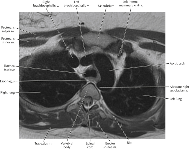
Chapter 9 Cardiac Anatomy Using MR AXIAL 2-CHAMBER 3-CHAMBER 4-CHAMBER SHORT AXIS Cardiac MR Axial 1 Normal Variant This axial MR image shows a common vascular variation—an aberrant right subclavian artery. Thoracic CT Soft Tissue Axial Plates 1 to 5 show the normal origin and course of the right subclavian artery. The aberrant right subclavian artery is one of the most common congenital vascular anomalies affecting the aortic arch and great vessels. Normally the right subclavian artery arises from the right brachiocephalic artery and courses into the right upper extremity (see Thoracic CT Soft Tissue Axial Plates 1 to 5). In contrast, the aberrant right subclavian artery typically arises from the distal aspect of the aortic arch or proximal descending thoracic aorta and courses through the mediastinum, posterior to the esophagus, to reach the right upper extremity. Cardiac MR Axial 2 Cardiac MR Axial 3 Cardiac MR Axial 4 Cardiac MR Axial 5 Technical Consideration These two MR images illustrate two of the many MR techniques used to examine the thoracic and cardiovascular system: “black blood” and “white blood” imaging techniques, named for the appearance of flowing blood on the image. There are a number of ways in which “black blood” images may be created, but these techniques have in common the lack of significant signal associated with flowing blood—hence, the vessels and cardiac chambers appear black. “White blood” sequences are often used for functional analysis and, although the bright signal within the vessels and cardiac chambers gives the impression that intravenous contrast has been injected, these imaging sequences do not require the use of intravenous contrast to generate signal within the vascular system. Cardiac MR Axial 6 Cardiac MR Axial 7 Diagnostic Consideration Only gold members can continue reading. Log In or Register to continue Share this: Share on X (Opens in new window) X Share on Facebook (Opens in new window) Facebook Related posts: Overview of Cardiac Anatomy Venous Anatomy and Variants Overview of Thoracic Anatomy Thoracic Lymph Nodes Thoracic Soft Tissue and Lung Cardiac Anatomy Using CT Stay updated, free articles. Join our Telegram channel Join Tags: Netters Correlative Imaging Cardiothoracic Anatomy Dec 26, 2015 | Posted by admin in CARDIOVASCULAR IMAGING | Comments Off on Cardiac Anatomy Using MR Full access? Get Clinical Tree