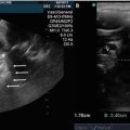
Fig. 3.1
Parasternal long-axis view. Visualization of the right ventricle (RV) superiorly, the left ventricle inferiorly, with the left atrium (LA) and the base or root of the aorta (aorta) on the left of the viewing screen. Also visualized are the left sided heart valves, i.e., the mitral and aortic valves
The parasternal long-axis (PLAX) view is the most comprehensive single view in the transthoracic echocardiography examination. With this one acoustic window, a qualitative, 2D functional assessment of both ventricles, the left-sided heart valves, and the aortic root can be obtained. The application of color Doppler further augments the interrogation of the mitral and aortic valves.
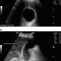
Short-Axis: Rotating the probe clockwise, (i.e., the plane of the ultrasound beam perpendicular to the plane of previously imaged long-axis view) allows visualizing of the short axis views of the heart. The transducer groove/notch should point superiorly, facing the right supraclavicular fossa, with the imaging beam parallel to an imaginary line joining the left shoulder and right flank.
In a “sweeping” fashion, the transducer is rotated in an anterior/caudal direction from a posterior/cranial angulation. In this manner, a cross section of the right ventricle and left ventricle, extending from the AV valves, through the middle of the ventricles, and out to apex, is obtained. Images are generated as if looking from the apex of the heart up toward the atria, i.e., LV is posterior and on the right, the RV is anterior and on the left (Fig. 3.2).


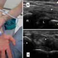
Fig. 3.2
Parasternal short axis views . Multiple short axis images through the right and left ventricles are obtained from the base of the heart (atrioventricular (AV) valve level) to the apex. (a) Base of the ventricles: The anterior and posterior mitral valve leaflets are seen in cross section (arrow). The right ventricle (RV) is anterior and on the right, the left ventricle (LV) is posterior and on the left. (b) Mid-ventricular level: The bodies of the anterolateral and posteromedial papillary muscles are visualized in cross section (arrow). (c) Apex: The apex of the right (RV) and left (LV) ventricles are displayed in cross section. The heads of the papillary muscle heads are not included in this view
The parasternal short-axis imaging is classically divided into three regions: (1) Base of the heart—the level of the mitral valve leaflets, (2) Mid-ventricle—the level of the anterolateral and posteromedial papillary muscles, and (3) Apex—distal to the papillary muscle head insertion. In a “Point of Care” environment, parasternal short axis imaging is instrumental in the rapid assessment for ventricular morphology and function.
Apical Imaging
Apical imaging is also obtained with the patient in the left lateral decubitus position. The transducer, however, is placed at or in the immediate vicinity of the point of maximal impulse (typically lateral and inferior to the left nipple). Three primary views are obtained from this position, all complimentary to the three short axis slices obtained from the parasternal windows, i.e., four-chamber view, apical long-axis view, and the two-chamber view. When combined, the apical long-axis and the parasternal short-axis images provide visualization of each segment of the left ventricle and therefore a complete view of left ventricular function.
Apical imaging display is arbitrary and determined via the physician acquiring the images, i.e., with the notch/groove of the transdu cer pointing upward, the left ventricle is on the right side, and with the notch/groove of the transducer pointing downward, the left ventricle is on the left side.
Four-Chamber View
With the ultrasound beam directed superiorly and medially (towards the patient’s right scapula), the ventricles, atria, interatrial and interventricular septa, and the AV valves are visualized in a single image. The apex of the left ventricular is positioned at the top of the image and the atria at the bottom. The goal of this image, via delicate manipulation the ultrasound beam anteriorly and posteriorly, is to visualize the atrial and ventricular septa in their entirety (Fig. 3.3). With regards to anatomic landmarks, the right-sided chambers are demarcated via the inferior insertion, by 5–10 mm, of the septal leaflet of the tricuspid valve relative to the anterior leaflet of the mitral valve on the left.

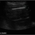
Fig. 3.3


Apical four-chamber view . As per standard echocardiographic convention, the left sided chambers of the heart are displayed on the left side of the imaging screen. The right (RV) and left (LV) ventricles, and the right (RA) and left (LA) atria are partitioned by the interventricular and interatrial septa, which join at the fibrous trigone of the atrioventricular (AV) valves to form the crux of the heart. The atrioventricular valves (mitral and tricuspid) are visualized as well; note the apical displacement, relative to the anterior mitral valve leaflet, of the septal leaflet of the tricuspid valve
Related posts:

Stay updated, free articles. Join our Telegram channel
Full access? Get Clinical Tree
