and Horia Ples2
(1)
Department of Computed Tomography, SCM Neuromed, Timisoara, Romania
(2)
Department of Neurosurgery, University of Medicine and Pharmacy “Victor Babes”, Timisoara, Romania
2.5 Carotid Angiography: Calcified Atheromatous Lesions and Kinking of Arteria Carotis Interna Sinistra
2.1 Normal Carotid Angiography
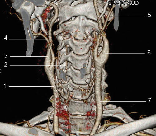
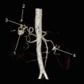
Fig. 2.1
Normal carotid angiography
1. A. carotis communis dextra
2. A. vertebralis dextra
3. A. carotis interna dextra
4. A. carotis externa dextra
5. A. carotis externa sinistra
6. A. carotis interna sinistra
7. A. carotis communis sinistra
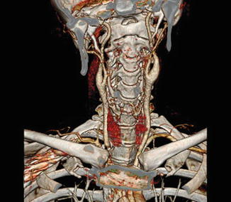
Fig. 2.2
Normal carotid angiography 3D VRT colour reconstruction anterior coronal plane
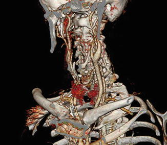
Fig. 2.3
Normal carotid angiography 3D VRT colour reconstruction oblique anterior plane
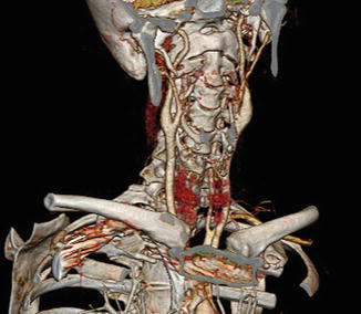
Fig. 2.4
Normal carotid angiography 3D VRT colour reconstruction left anterior oblique plane
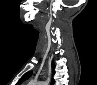
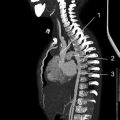
Fig. 2.5
Normal carotid angiography 3D MIP reconstruction with visualisation of a. carotis communis sinistra and a. carotis interna sinistra
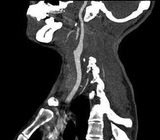
Fig. 2.6
Normal carotid angiography 3D MIP reconstruction with visualisation of a. carotis communis dextra and a. carotis interna dextra
2.2 Anomalous Origin of Arteria Carotis Communis
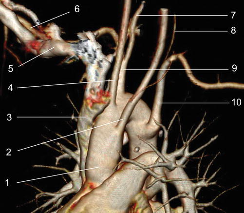
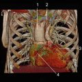
Fig. 2.7
Anomalous origin of common carotid arteries
1. Aorta ascendens
2. A. carotis communis sinistra
3. Vena cava superior
4. A. carotis communis dextra
5. V. subclavia dextra
6. A. subclavia dextra
7. A. vertebralis dextra
8. A. vertebralis sinistra
9. Truncus brachiocephalicus
10. A. subclavia sinistra
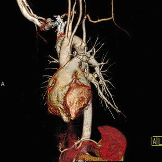
Fig. 2.8
Colour reconstruction 3D VRT
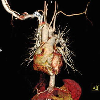
Fig. 2.9
Colour reconstruction 3D VRT
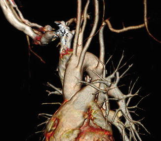
Fig. 2.10
Colour reconstruction 3D VRT, enlarged image, anterior plane
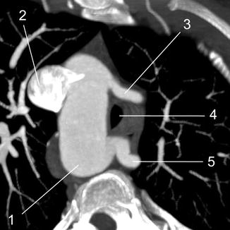
Fig. 2.11
Axial image, 3D MIP
1. Arcus aortae
2. Vena cava superior
3. A. carotis communis sinistra
4. Trachea
5. A. subclavia sinistra with retrotracheal and retro-oesophageal trajectory
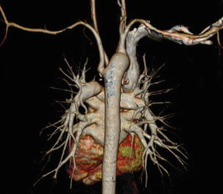
Fig. 2.12
3D VRT reconstruction, posterior plane
2.3 Calcified Atheromatous Plaques at the Level of Arteria Carotis
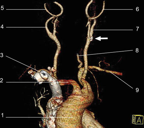
Fig. 2.13
Normal carotid angiography. The arrow indicate calcified plaques
1. Aorta ascendens
2. Truncus brachiocephalicus
3. V. cava superior
4. A. carotis communis dextra
5. A. carotis externa dextra
6. A. carotis externa sinistra
7. A. carotis interna sinistra
8. A. vertebralis sinistra
9. A. subclavia sinistra
10. A. carotis communis sinistra
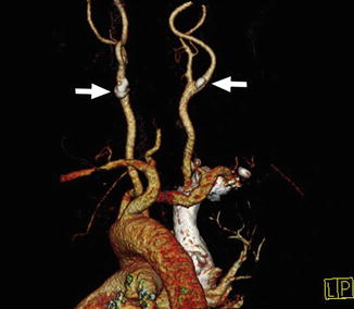
Fig. 2.14
3D VRT colour reconstruction, posterior plane. The arrow indicate calcified plaques
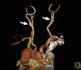
Fig. 2.15
3D VRT colour reconstruction, posterior plane. The arrows indicate calcified atheromatous plaques
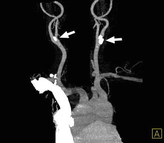
Fig. 2.16
3D VRT reconstruction
The arrows indicate calcified atheromatous plaques placed at the level of the emerging a. carotis interna
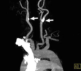
Fig. 2.17
3D VRT reconstruction
The arrows indicate calcified atheromatous plaques placed at the level of the emerging a. carotis interna
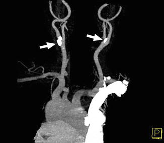
Fig. 2.18
3D VRT reconstruction
The arrows indicate calcified atheromatous plaques placed at the level of the emerging a. carotis interna
2.4 Carotid Angiography: Nonobstructive Calcified Atheromatous Plaques
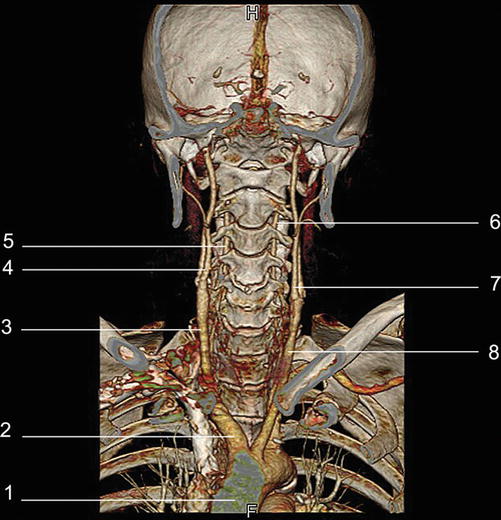
Fig. 2.19
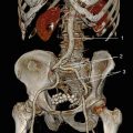
Reconstruction 3D VRT colour, frontal plane
1. Aorta ascendens
2. Truncus brachiocephalicus
3. A. carotis communis dextra
4. A. carotis interna dextra
5. A. vertebralis dextra
6. A. vertebralis sinistra
7. A. carotis interna sinistra
8. A. vertebralis sinistra
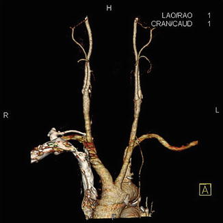
Fig. 2.20
3D VRT colour reconstruction, after removal of the bone structure
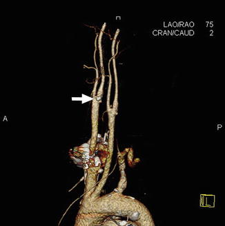
Fig. 2.21
3D VRT colour reconstruction, oblique and sagital planes
The arrows indicate calcified atheromatous plaques
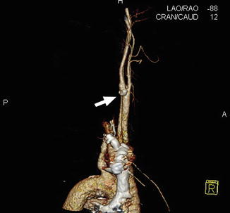
Fig. 2.22
3D VRT colour reconstruction, oblique and sagital planes
The arrows indicate calcified atheromatous plaques
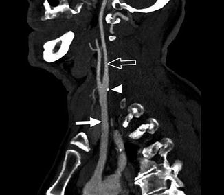
Fig. 2.23
3D MIP reconstruction
Full arrow = a. carotis communis sinistra
Arrow with contour = a. carotis interna sinistra
Tip of the arrow = atheromatous calcified plaque
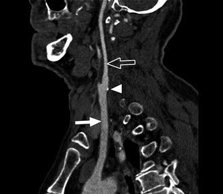
Fig. 2.24
3D MPR reconstruction
Full arrow = a. carotis communis sinistra
Arrow with contour = a. carotis interna sinistra
Tip of the arrow = atheromatous calcified plaque
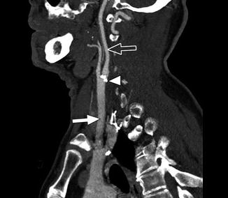
Fig. 2.25
3D MIP reconstruction
Full arrow = a. carotis communis dextra
Arrow with contour = a. carotis interna dextra
Tip of the arrow = atheromatous calcified plaque
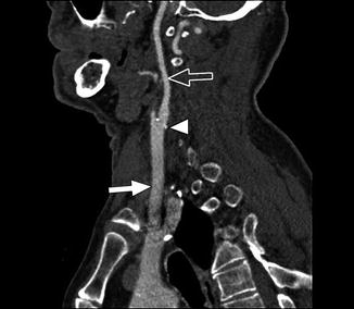
Fig. 2.26


3D MPR reconstruction
Full arrow = a. carotis communis dextra
Arrow with contour = a. carotis interna dextra
Related posts:
Stay updated, free articles. Join our Telegram channel
Full access? Get Clinical Tree
