Cecal Volvulus
Shaun R. Rybak
CLINICAL HISTORY
48-year-old female with acute abdominal pain and bloating.
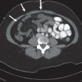
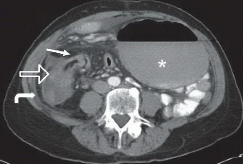 Figure 23A |
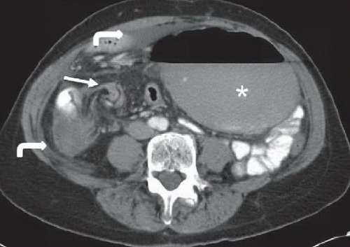 Figure 23B |
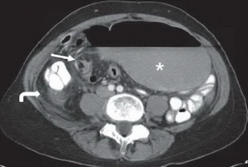 Figure 23C |
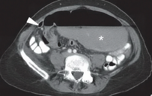 Figure 23D |
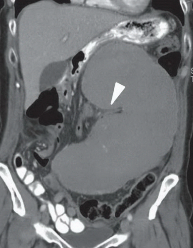 Figure 23E |
FINDINGS
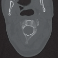
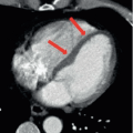
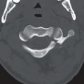
Figures 23A, 23B, 23C, and 23D: Axial contrast-enhanced CT images of the abdomen demonstrate a dilated volvulated cecum (asterisk) with a long air-fluid level and whirling/twisting of the mesenteric vessels and colon in the right lower quadrant (arrow). The right colon is decompressed (open arrow in Fig. 23A). Small volume of free fluid is noted on the axial images (curved arrow). Figure 23D best
shows the beaking of the cecum as it begins twisting on itself (arrowhead). Figure 23E: Coronal CT image of the abdomen shows the kidney bean shape of the dilated volvulated cecum, which points to the left upper quadrant. The dilated cecum bends on itself at the level of the terminal ileum and ileocecal valve (arrowhead).
shows the beaking of the cecum as it begins twisting on itself (arrowhead). Figure 23E: Coronal CT image of the abdomen shows the kidney bean shape of the dilated volvulated cecum, which points to the left upper quadrant. The dilated cecum bends on itself at the level of the terminal ileum and ileocecal valve (arrowhead).
Related posts:


Stay updated, free articles. Join our Telegram channel

Full access? Get Clinical Tree


