, Foster D. Lasley2, Indra J. Das2, Marc S. Mendonca2 and Joseph R. Dynlacht2
(1)
Department of Radiation Oncology, CHRISTUS St. Patrick Regional Cancer Center, Lake Charles, LA, USA
(2)
Department of Radiation Oncology, Indiana University School of Medicine, Indianapolis, IN, USA
Cell and Tissue Kinetics: Why Do We Care?
Redistribution and Repopulation are two of the four “R’s” of radiobiology (below).
Redistribution between different parts of the cell cycle can cause different effects depending on dose-rate and fractionation.
Repopulation simply increases cell survival during a protracted course of treatment. Some tumors and normal tissues repopulate faster than others.
The “4 R’S” of Radiobiology
Repair
Reoxygenation
Redistribution
Repopulation
The “fifth R” of Radiosensitivity
Definitions
G 1 : Gap phase 1, DNA is un-duplicated.
S: Synthesis phase, DNA is being duplicated.
G 2 : Gap phase 2, DNA is fully duplicated.
M: Mitosis phase, chromosomes condense, the nucleus and cell divides.
T C : Cell cycle time (total duration of all phases)
T G1 : G1 phase duration
T S : S phase duration
T G2 : G2 phase duration
T M : M phase duration
MI: Mitotic index
LI: Labeling index
λ: Cell distribution correction factor
T vol : Tumor volume doubling time
T d : Tumor diameter doubling time
T pot : Potential doubling time of tumor
GF: Growth fraction
CLF (ϕ): Cell loss factor
Molecular Biology of the Cell Cycle
The cell must pass through checkpoints to continue through the cell cycle.
Each checkpoint is governed by Cdk–Cyclin complexes and their inhibitors.
These regulatory molecules change the amount and activity of other proteins, leading to cell cycle progression.
G 1 /S: Cyclin D, Cdk4/6
G 1 /S: Cyclin E, Cdk2
S: Cyclin A, Cdk2
G 2 /M: Cyclin B, Cdk1 (Fig. 24.1).
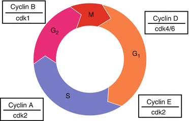
Fig. 24.1
The cell cycle. The cell has checkpoints regulated by the Cdk-Cyclin complexes that respond to other signals in the cell.
The G 1 /S checkpoint is frequently inactivated in human tumors:
The Cyclin D/cdk4/6 complex is inhibited by p21 and p15/p16 (INK4A).
Mitogenic signals cause Cyclin D activation.
The activated Cyclin D/cdk4/6 complex partially phosphorylates and activates the retinoblastoma tumor suppressor Rb which leads to release of the E2F transcription factor family.
Release of E2F, leads to transcription of Cyclins E and A, and genes involved in DNA synthesis.
Cyclin E/Cdk2 and Cyclin A/Cdk2 complexes further phosphorylates Rb, leading to transition into and through S phase.
p53 blocks the G 1 /S transition by induction of p21 in response to DNA damage or other cellular stress.
Rb or p53 are frequently defective in tumors. They may be directly mutated, or may be inhibited by other pathways.
This allows for uncontrolled entry into S phase.
The G2/M cell cycle transition is regulated by the CyclinB/Cdk1 complex which phosphorylates histone H1 and nuclear lamins leading to chromosome condensation and dissolving of the nuclear membrane in preparation for mitosis.
The G2/M checkpoint can also be induced by p21 binding.
It is important to remember that the G2/M checkpoint remains intact in most cancers.
Imaging the Cell Cycle
Light microscopy:
Can detect M-phase cells by morphology and chromosome condensation.
Cannot tell the difference between G 1 , S and G 2 .
Mitotic Index (MI) = % of cells in M phase.
3 H-Thymidine:
Tritium ( 3 H) is a low energy (19 keV) beta emitter commonly used in the laboratory. It is detected by autoradiography.
Culturing cells in 3H-Thymidine will label cells in S-phase as it is readily incorporated in replicating DNA.
Labeling Index (LI) = % of cells in S phase.
Microscopy and autoradiography of 3 H-Thymidine labeled cells can be used to measure MI and LI simultaneously.
Cell Cycle Kinetics: Measuring TM and TS (Fig. 24.2)
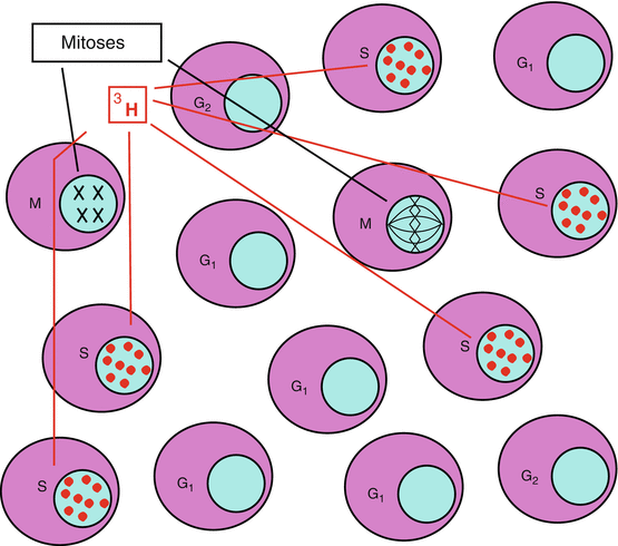
Fig. 24.2
Light microscopy can be used to view cells in mitosis and therefore measure the mitotic index. The addition of tritiated thymidine allows cells in S-phase to be imaged by autoradiography, permitting measurement of the labeling index.
Stay updated, free articles. Join our Telegram channel

Full access? Get Clinical Tree


