Cerebral Contusions
Benjamin Y. Huang
CLINICAL HISTORY
63-year-old female who presents to the emergency room with headache and nausea following a syncopal episode.
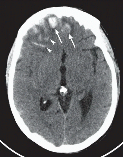 FIGURE 37A |
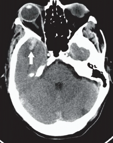 FIGURE 37B |
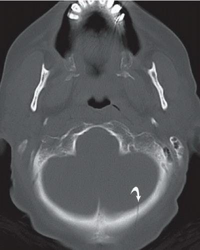 FIGURE 37C |
FINDINGS
Figures 37A and 37B: Axial noncontrast head CT images displayed in brain windows demonstrate hyperdense, hemorrhagic intra-axial lesions involving cortex and subcortical white matter with mild surrounding edema, situated immediately subjacent to the inner cortex of the calvarium in the most anterior portions of the bilateral frontal lobes (arrows in Fig. 37A) and right temporal lobe (wide arrow in Fig. 37B). There is also evidence of subarachnoid hemorrhage in the right frontal lobe sulci (arrowhead in Fig. 37A). Image through the posterior fossa displayed with a bone window (Fig. 37C) demonstrates a nondisplaced left occipital bone fracture (curved arrow).
Related posts:


Stay updated, free articles. Join our Telegram channel

Full access? Get Clinical Tree


