CERVICOTHORACIC JUNCTION: BRACHIAL PLEXUS VASCULAR CONDITIONS
KEY POINTS
- Brachial plexus/cervicothoracic junction vascular abnormalities may mimic other conditions.
- These abnormalities may pose an immediate functional risk to the patient.
- Imaging can most often provide a very likely diagnosis and establish whether the process is primarily or secondarily affecting the vasculature and brachial plexus.
- Imaging can rapidly exclude significant structural pathology with a high degree of confidence.
- Imaging in potential cervicothoracic junction vascular problems is pivotal in proper medical triage and further decision making.
Vascular conditions that involve the cervicothoracic junction (CTJ) and brachial plexus are relatively uncommon. They are a possible source of symptoms or even a mass lesion, and a vascular etiology must be considered when a mass is accompanied by signs and symptoms of ischemia or venous occlusion. Such conditions are often lumped under the term thoracic outlet syndrome (TOS); however, TOS, in the vast majority, manifests only neurologically in the form of a brachial plexopathy.
Other vascular conditions that manifest at the CTJ are considered here for completeness. These include vascular malformations, vasculitis, dissection, true aneurysms, and contained leaks as well as venous thrombosis and thrombophlebitis.
ANATOMIC CONSIDERATIONS
Applied Anatomy: Cervicothoracic Junction and Brachial Plexus
The overall anatomy of the CTJ and brachial plexus is discussed in Chapter 149 for the infrahyoid neck in general and more specifically in Chapter 163 for the CTJ region. While the specific anatomy that follows must be considered in all patients with brachial plexopathy, it is considered here because of its possible links to compressive vascular occlusion in patients with TOS that might be approached with a surgical decompression (Fig. 165.1).
The trunks of the brachial plexus and subclavian vessels may be compressed or irritated where they pass within three narrow regions from the low neck and CTJ to the axilla. The first, and most critical, of these regions is the most proximal interscalene triangle, whose borders are the anterior scalene muscle anteriorly, the middle scalene muscle posteriorly, and the medial surface of the first rib inferiorly. Fibrous bands, cervical ribs, and anomalous muscles may narrow this already tight triangle.
The second region of possible constriction is the costoclavicular triangle, which is bordered by the middle third of the clavicle anteriorly, by the first rib posteriorly and medially, and by the upper border of the scapula posteriorly and laterally.
The third and most distal region of possible constriction is the subcoracoid space beneath the coracoid process lying deep to the pectoralis minor tendon.
IMAGING APPROACH
Techniques and Relevant Aspects and Imaging Pros and Cons
These are discussed in Chapter 149 for the infrahyoid neck in general and more specifically in Chapter 163 for the CTJ and are presented in Appendixes A and B.

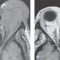
FIGURE 165.1. A patient, originally diagnosed as having vasculitis, presenting with supraclavicular fullness and tenderness and symptoms suggesting vascular insufficiency in the right arm. A: Contrast-enhanced T1-weighted (T1W) image showing enhancement within and around the scalene muscle group (arrows) but is otherwise not definitive. B: Contrast-enhanced T1W coronal image showing the subclavian artery wall to enhance (arrows) and that the lumen is potentially patent. C, D: Flow-sensitive gradient echo image showing that the right subclavian artery is actually clotted. That finding was confirmed on the maximum intensity projection (MIP) image in (D). (NOTE: The patient was treated for the acute vascular occlusion and then decompressed for compressive vascular thoracic outlet syndrome.)
Stay updated, free articles. Join our Telegram channel

Full access? Get Clinical Tree


