Challenge

Challenge


Case 157


Comment


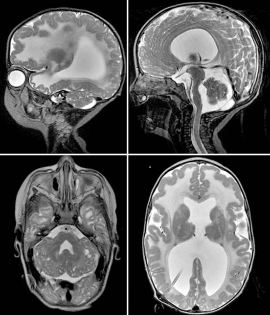
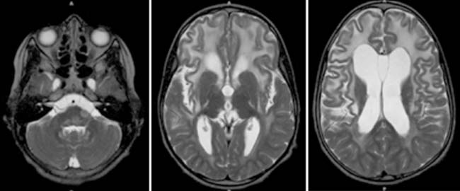
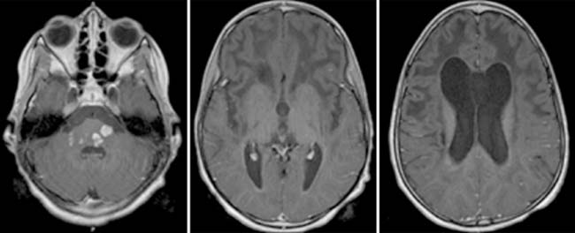
MEB belongs to the group of CMDs, which is a heterogeneous group of congenital, hereditary myopathies that are often progressive. Frequently, the name of the diagnosis is based on the combined abnormalities of the eyes and brain. MEB is inherited autosomal recessive. Early clinical symptoms include muscular hypotonia and poor visual contact. A hydrocephalus is seen in the majority of patients. Motor development is generally severely delayed. In addition, severe mental retardation is usually present in combination with seizures. Ocular signs include severe myopia, glaucoma, retinal degeneration, choroidal hypoplasia, and optic nerve hypoplasia. MEB is caused by a defect in the O-mannosyl glycan synthesis. No curative treatment is available.


MRI findings are extensive and prominent. Ventriculomegaly frequently ranges from mild-to-significantly dilated. Additional striking features include a hypoplastic pons and brainstem, multiple subcortical cerebellar cysts, and variable degrees of cerebral polymicrogyria. The cortex is characteristically thickened, and the inner contour of the cortex reveals the polymicrogyria most prominently. In addition, the cerebral white matter is edematous and swollen with a significantly increased T2-weighted signal. The mesencephalon is frequently elongated with a malformation of the tectal plate. The superior and inferior colliculi may be fused. In addition, neuronal heterotopias may be seen within the cerebral hemispheres, as well as a thinned, hypoplastic corpus callosum and a partial absence of the septum pellucidum. The characteristic features, especially the combination of a ventriculomegaly and a hypoplastic, elongated, and kinked brainstem may already be observed on prenatal, fetal MRI. The cerebellar cysts may, however, be too small to be detected intrauterine or may progress after birth.


Case 158


Comment


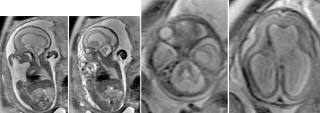
Rhombencephalosynapsis is a complex congenital malformation of the posterior fossa, characterized by a fusion of the cerebellar hemispheres, dentate nuclei, and hypoplastic or fused superior cerebellar peduncles (single-lobed cerebellum). In addition, the cerebellar vermis is absent or hypoplastic. This malformation is believed to be the result of a defective dorsal induction or a differentiation of the cerebellar midline structures or both. Rhombencephalosynapsis has been linked to several gene defects, but it is also observed in conditions such as Gomez-Lopez-Hernandez syndrome. Rhombencephalosynapsis is extremely rare; the clinical presentation is usually nonspecific with ataxia, gait abnormalities, and developmental delays. Associated findings, such as corpus callosum agenesis or dysgenesis, septo-optic dysplasia, and hydrocephalus as a result of aqueductal stenosis, are frequently observed. Migrational abnormalities with cerebral polymicrogyria have also been described. Rhombencephalosynapsis may be diagnosed intrauterine by fetal magnetic resonance imaging (MRI) or by postnatal MRI. Imaging findings are usually obvious with the identification of a single-lobed cerebellum with folia and cerebellar fissures crossing the midline (transverse folia). In addition, the dentate nuclei are fused and usually in a more midline position dorsally to a narrowed or keyhole-shaped fourth ventricle. Midsagittal images may be misleading; however, the pattern of the midline fissures do not match the normal anatomy of the vermis (i.e., the primary fissure is missing). Coronal and axial images are most useful. The prognosis is variable; most children do not survive until adulthood. Additional lesions or complications such as hydrocephalus or both shorten survival.


Case 159


Comment


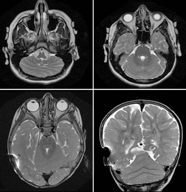
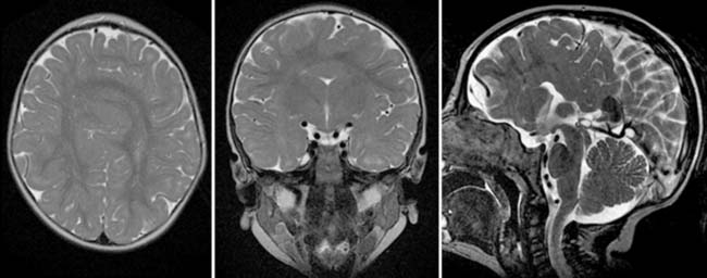
Joubert syndrome was originally reported by Dr. Marie Joubert who described several children with a similar clinical presentation that included episodic hyperpnea, abnormal eye movement, rhythmic tongue protrusions, ataxia, and mental retardation. Anatomically, the cerebellar vermis is lacking. On MRI, the axial images show a characteristic molar tooth appearance of the brainstem, resulting from a horizontal course of the thickened superior cerebellar peduncles. The fourth ventricle is deformed and resembles an umbrella or bat wing on axial planes. Because the vermis is lacking, both cerebellar hemispheres reach each other in the midline; consequently, the midline slices reveal cerebellar fissures on sagittal imaging instead of vermian sulci. In addition, dentate nuclei are lateralized. Tractography studies have shown that the corticospinal tracts do not cross at the pyramidal decussation, which may, in part, explain the clinical presentation. Multiple gene loci that are linked to Joubert syndrome have been identified. The expression may vary; children with Joubert syndrome may show different degrees of mental retardation and gait disturbance. Currently, no prenatal test is available for early diagnosis. Rarely associated supratentorial anomalies occur, and cortical dysplasia and gray-matter heterotopia have been reported. In children with Joubert syndrome, the kidneys may be affected (cerebelloocculorenal syndrome). Renal ultrasound should be considered during the diagnostic work-up of these children. Experienced pediatric neurologists may suspect a diagnosis, based on the facial characteristics that are not specific but indicative. With ongoing research, many more malformations of the brainstem and cerebellum that share a molar tooth appearance of the brainstem are described. Perhaps, Joubert syndrome is simply one of a spectrum of brainstem malformations.


Case 160


Comment


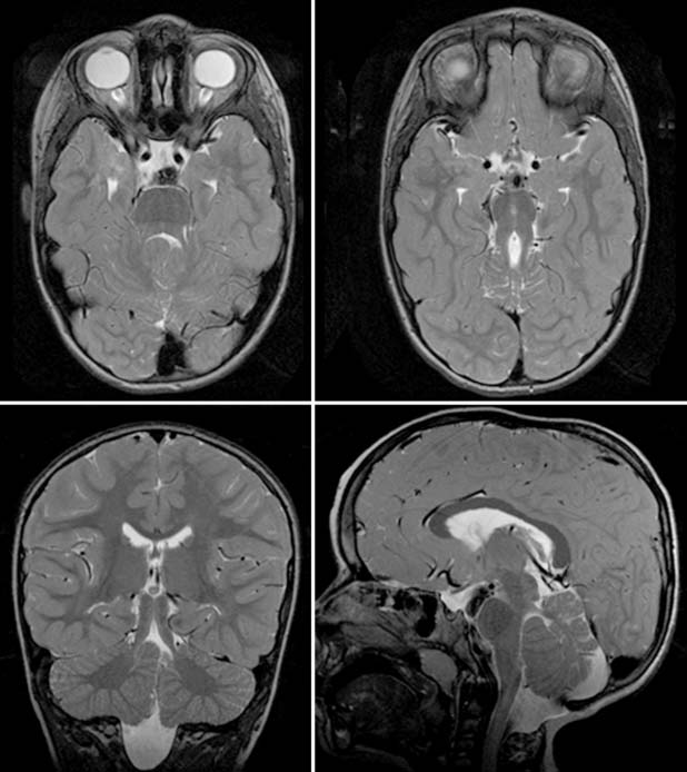
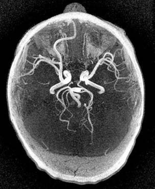
A group of holoprosencephalies results from a disorder of diverticulation or cleavage. Holoprosencephalies are also known as disorders of ventral induction. Holoprosencephalies encompass an entire spectrum of malformations; the mildest form is septo-optic dysplasia; the most severe form is alobar holoprosencephaly. They both result from a failure of cleavage of the prosencephalic vesicle during organogenesis. Frequently, associated anomalies of the face are seen—“The face predicts the brain.” Cyclopia has been described in cases of alobar holoprosencephaly. Clinically, depending on the severity of the malformation, different symptoms may occur and include seizures, mental retardation, dystonia, microcephaly, hypothalamic-pituitary dysfunction, cyclopia, and fused metopic suture. Symptoms may be mild in lobar holoprosencephaly and severe in alobar holoprosencephaly. In septo-optic dysplasia, the anterior horns of the ventricles have a boxlike configuration on coronal MRI. In addition, an optic nerve pathologic abnormality is seen on fundoscopy. Various degrees of hypothalamic-pituitary dysfunction are observed. In the lobar holoprosencephaly, a lobar brain is seen with hypoplastic frontal lobes, some frontal horn formation, and the falx cerebri extends frontally. In semilobar holoprosencephaly, a partially formed fax cerebri and a partially formed interhemispheric fissure are seen in the posterior part. The anterior brain is fused, the thalami are partially separated, and a small third ventricle and rudimentary temporal horns are demonstrated. The septum pellucidum is absent, the splenium of the corpus callosum is present, whereas the truncus of the corpus callosum is lacking. Hypoplastic olfactory bulbs and optic nerves may exist. In alobar holoprosencephaly, a small holosphere and monoventricle are seen, as well as fused thalami. However, no third ventricle, falx cerebri or corpus callosum, or interhemispheric fissure is seen. In addition, no temporal horns are demonstrated, and malformations of the Willis circle are observed with an azygos or unpaired anterior cerebral artery. The prognosis is frequently poor, especially in the most severe forms of holoprosencephalies. Diagnosis is usually made prenatally by either ultrasound or fetal MRI. In septo-optic dysplasia, the prognosis will be determined by the associated malformations. In up to 50% of cases, additional lesions are identified by MRI. Postnatally, MRI is indicated to identify all details of the malformation.


Case 161


Comment


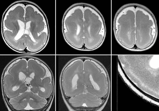
Neuronal migration anomalies and malformations of cortical development occur between the second and fourth months gestation. In neuronal migration anomalies, cells that should migrate from the periventricular germinal matrix to the cortical surface of the brain are arrested somewhere along their paths. These cells may rest at multiple locations, either focal or as bands. Heterotopic gray matter may be recognized on imaging focally as subependymal heteroptopic cell masses or as bands within the periventricular, central, or subcortical white matter of the cerebral hemispheres. Disorders of cortical organization may result in agyria, pachygyria, polymicrogyria, or focal cortical dysplasia. Frequently, combined malformations of neuronal migration and cortical organization are seen. In lissencephaly (type I), an extensive arrest of the neuronal migration results in a wide, T2-hypointense band of neurons within the white matter of both hemispheres separated from the overlying cortex by a thin band of T2-hyperintense white matter. The overlying cortex is typically lissencephalic and has a four-layer architecture combined with a smooth brain surface. The anomaly is usually symmetric. Ventriculomegaly may be associated. The cerebellum is usually unremarkable. Children may present with intractable seizures and a developmental delay. The cause is not yet fully determined but is at least known to be mediated genetically but is also believed to be the result of intrauterine infections. MRI is the imaging modality of choice to identify the exact anatomy of the anomaly. High-resolution imaging will also display that the subcortical white matter has multiple linear bands of T2-hypointensity, which most likely represents streaks of neurons that almost reached the lissencephalic cortex. Prenatal diagnosis on ultrasound may be difficult because the overlying fetal skull prevents a detailed study of the cortex; the arrested neurons within the white matter are also difficult to detect. Lissencephaly (type I) should be differentiated from the neuronal migration anomalies and malformations of cortical development because differentiation will help determine prognosis.


Case 162


Discussion


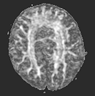
PML is a severe, demyelinating disease of the central nervous system as a result of JC papovavirus infection of the myelin-producing oligodendrocytes. The name JC virus is derived from the initials of the index patient. PML typically occurs in immunocompromised individuals, such as children with congenital HIV infection or other conditions associated with impaired T-cell function. In the course of the infection, extensive myelin breakdown results in white matter destruction. Neurologic symptoms are unspecific and include focal neurologic deficits and dementia. Without treatment, patients have a relentless downhill course. The disease is usually fatal within 1 year of diagnosis in 90% of patients. The diagnosis can be established by the detection of the JC virus deoxyribonucleic acid (DNA) in cerebrospinal fluid by polymerase chain reaction testing. Many reports have described MRI findings in patients with PML, but only few MRI abnormalities correlate with patient survival. Conventional MRI reveals patchy areas of T2-hyperintense and T1-hypointense demyelination within the white matter. These lesions do not usually show any contrast enhancement and have minimal or no mass effect. Typically, these lesions are asymmetric. On diffusion tensor imaging, the lesions have a reduced fractional anisotropy, confirming the injury to the white matter by the infection. On follow-up imaging, lesions may show a rapid increase without treatment. The overlying cortex may be thinned. Any child with a rapidly expanding, focal white matter lesion with a compromised immune system should be suspected for PML until proved otherwise. The differential diagnoses may include acute disseminated encephalomyelitis and radiation necrosis or metabolic white matter diseases. The rapid progression and the clinical history are usually helpful in differentiating the cause of the observed lesions.


Case 163


Comment


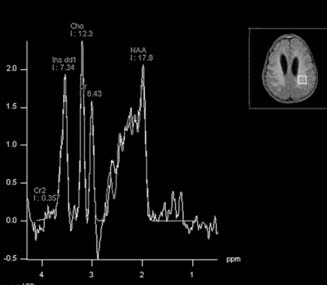
Alexander disease is a leukodystrophy first described by W. Stewart Alexander in 1949 as progressive fibrinoid degeneration of fibrillary astrocytes associated with mental retardation in a hydrocephalic infant. Three subgroups have been identified: infantile, juvenile, and adult. In the juvenile subgroup, the patients become symptomatic between 4 and 14 years of age, typically with a progressive loss of previously mastered developmental milestones. In addition, patients may present with progressive bulbar and pseudobulbar symptoms, an increase in swallowing problems and apneic attacks, impaired speech or loss of speech, dysarthria, hoarseness, spasticity, cerebellar ataxia, seizures, and progressive behavioral and cognitive deterioration. Average duration of the illness is 8 years. Most cases are sporadic, but familial cases have been described. Imaging features are rather characteristic with predominant frontal lobe involvement that may progress over the course of the disease into the parietal regions. Five characteristic MRI criteria have been defined, and four of the five have to be fulfilled for diagnosis: (1) symmetric cerebral white matter abnormalities with predominant frontal involvement, (2) T2-hypointense, T1-hyperintense rim of tissue along the ventricles, (3) focal lesions in the basal ganglia and thalami, (4) brainstem abnormalities (midbrain and medulla), and (5) contrast enhancement of the described lesions including optic chiasm. In addition, 1H-MRS will show increased myoinositol and choline levels within the affected white matter, as well as a reduced N-acetyl aspartate peak. Lactate may also be observed. On microscopic examination, countless Rosenthal fibers are seen throughout the brain within hypertrophic fibrillary astrocytes. Macrocephaly results from the combination of astrocytic hyperplasia and massive deposition of Rosenthal fibers.


Case 164


Case 165


Comment


Thyroglossal duct cysts are the most common congenital cysts in the neck representing 75% of midline masses and resulting from cystic dilation of epithelial remnants of the thyroglossal duct tract, which is present during the embryologic inferior migration of the thyroid from the base of the tongue to its normal final location. These cysts present as midline neck masses at the level of the thyrohyoid membrane and are closely associated with the hyoid bone. Most patients present as children, although presentation at any age is possible. Males and females are equally affected, and the cysts are usually asymptomatic, but they may become infected and form abscesses and draining fistulas.


In this example, the cyst is small; if larger, both an elevation of the tongue and a swallowing dysfunction would have developed. Ultrasound is the imaging modality of choice because it is ideal for determination of the nature of the mass and the localization of thyroid tissue. In this case, the lesion appears as a complex cyst, which is likely because of prior infection or hemorrhage. Computed tomography (CT) and magnetic resonance imaging (MRI) is also diagnostic but radiation (CT), length of study (MRI), and expense make both modalities impractical and unnecessary. Scintigraphy may be helpful to localize ectopic location of the thyroid gland.


Surgical resection is considered the treatment of choice. If the patient is euthyroid and thyroid tissue is seen in the normal location, no further imaging of the thyroid gland is necessary. In cases of abnormal thyroid function, nuclear thyroid scans are indicated to search for ectopic thyroid. Ectopic thyroid tissue may be located in the sublingual space in 10% of patients and at the base of the tongue in 90% of patients. In 75% of patients, the ectopic tissue is the only functioning thyroid tissue. However, these patients are usually hypothyroid anyway because the ectopic tissue is dysplastic. A careful histologic study of the specimen is important; a small incidence of carcinoma arising in thyroglossal duct remnants exists.


Case 166


Case 167


Comment


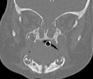
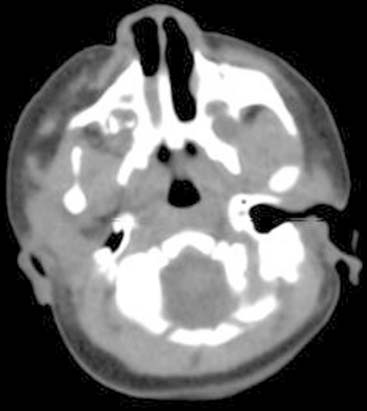
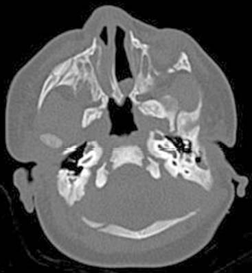
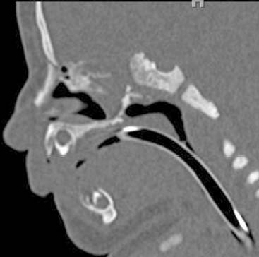
Newborns are obligatory nasal breathers; therefore nasal obstruction is an airway emergency in this population. Choanal atresia is the result of an occluded nasal airway and typically presents with respiratory distress during feeding, which may be alleviated by crying. Theories proposed to explain the embryogenesis of choanal atresia include persistence of the buccopharyngeal membrane and the oronasal membrane, as well as the mesodermal flow theory, in which a misdirection of neural crest cell migration, secondary to local factors, is implicated. Bilateral congenital choanal atresia should be suspected when the infant presents signs of asphyxia and cyanosis that improves with crying. Choanal atresia will only present as an airway emergency if it is bilateral. Choanal atresia occurs in 1 of 8000 live births and is commonly (>75%) associated with syndromes and systemic anomalies. Approximately two thirds of patients are bilateral. In the most severe form of this condition, the hard palate and vomer are fused with the ventral clivus, and the nasopharynx is very small (nasopharyngeal atresia). In another form, membranous choanal atresia is a result of the failure of the buccopharyngeal membrane to perforate. In more than 90% of patients with choanal atresia, the abnormality is partly or completely osseous; pure membranous atresia is rare. Female infants are affected twice as often as male newborns. Computed tomography is the imaging modality of choice to diagnose and help determine whether the obstruction is bony or membranous. Multiplanar reconstructions should be performed to better evaluate the anatomy. Normal choanal orifices measure more than 0.37 cm in children younger than 2 years of age. The width of the posterior and inferior parts of the vomer is normally less than 0.34 cm in patients younger than 8 years. Additional congenital anomalies are present in approximately 50% of the patients and should be actively searched for in the presence of choanal atresia.


Case 168


Case 169


Comment


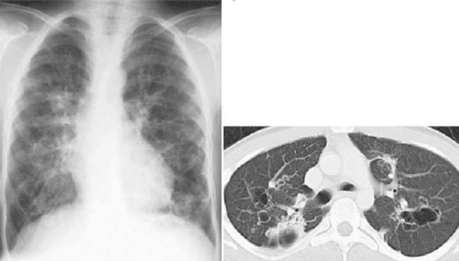
Recurrent respiratory papillomatosis (RRP) is relatively rare in children. However, papillomas are the most common tumor found in the larynx in childhood.


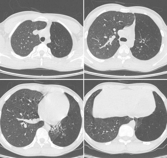
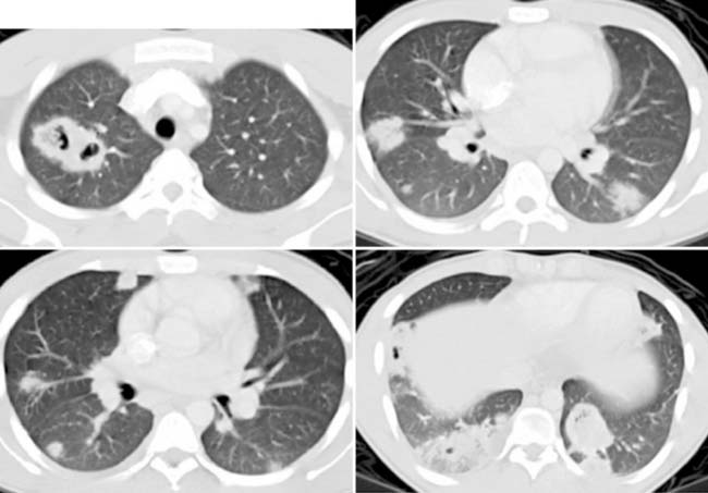
Benign tumors of the aerodigestive tract from the human papilloma viral infection result in papillomatosis. Lesions may be 1 mm to several centimeters in size. The most frequent location is the larynx (95%) with the tracheobronchial tree, lung parenchyma, nasopharynx, and oropharynx and esophagus less common. Endobronchial spread leads to lung nodules that may cavitate. Complications include airway obstruction, hemorrhage, and rarely malignant degeneration.


Hoarseness is the most common clinical presentation, but it may also include a weak cry, stridor, and failure to thrive. Most patients present between the ages of 2 and 5 years. At least one half of affected children have mothers with condyloma acuminata, but most infants of mothers with genital papilloma viral infection do not develop RRP.


Diagnosis is made with laryngoscopy. Most cases can be treated with laser therapy, but lesions frequently recur.


Laryngeal tracheopapillomatosis spreads to the lungs in less than 1% of patients and is associated with a poor prognosis. Radiographic findings include thin-walled cystic lesions, as well as solid pulmonary lesions that tend to grow peripherally and lead to lung destruction. Lesions are distributed predominantly in the posterior lower lobes and are frequently associated with atelectasis, bronchiectasis, and associated infections. Differential diagnoses include granulomatous disease (Wegener disease), metastatic disease, and septic emboli.


Case 170


Comment


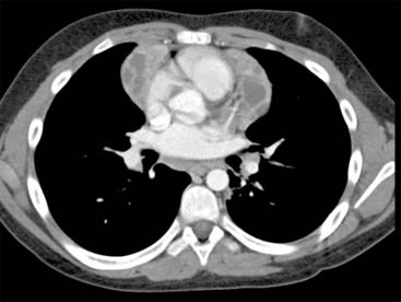
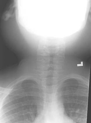
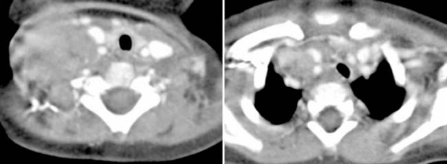
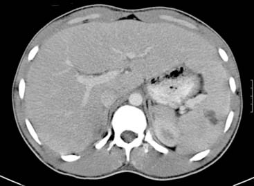
Lemierre syndrome is pharyngitis with septic thrombosis of the internal jugular veins and is usually caused by the bacterium Fusobacterium necrophorum. It typically affects young, previously healthy patients. Lemierre syndrome develops most often after oropharyngeal infection; the pathogens spread to the internal jugular vein by several anatomic routes including local draining veins, lymphatic tissue, or direct extension through the soft tissue of the neck. The bacteria penetrate the jugular vein, causing an infected clot, from which bacteria are seeded throughout the body. Pieces of the infected clot break off and travel to the lungs as emboli, blocking branches of the pulmonary artery. If untreated, supportive infection can spread directly into the ear, mediastinum, and cranial vault. Findings on clinical examination of patients with Lemierre syndrome are quite variable. Signs of exudative tonsillitis, pharyngitis, and thick gray pseudomembranes, as well as oral ulcers, have been reported. However, oropharyngeal examination may be normal because sepsis typically occurs 1 week after primary infection, allowing time for physical findings to resolve. Neck pain, swelling, and trismus along the anterior border of the sternocleidomastoid muscle and at the angle of the mandible are the clinical manifestations of a septic thrombophlebitis of the ipsilateral internal jugular vein. Signs and symptoms of pulmonary embolism may be the presenting clinical finding, especially in patients in whom oropharyngeal manifestations have resolved. Persistent fever and rigors despite antibiotics, followed by the complaint of pleuritic chest pain, hemoptysis, or dyspnea, may be all that is required for a presumptive diagnosis.


Case 171


Comment


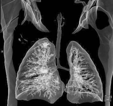
Between 1% and 12% of patients who undergo bronchography or bronchoscopy demonstrate some form of congenital tracheobronchial variant or anomaly. Contrary to the numerous variations of lobar or segmental bronchial subdivisions, abnormal bronchi originating from the trachea or main bronchi are rare.


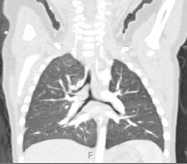
Tracheal bronchus was first described in 1785 as an airway malformation in which the right upper lobe bronchus originates in the trachea. The term tracheal bronchus includes a variety of bronchial anomalies that affect the trachea or main bronchus and are directed toward the upper lobe territory. This anomalous bronchus usually exits the right lateral wall of the trachea less than 2 cm above the major carina and can supply the entire upper lobe or its apical segment. The two types are (1) displaced and (2) supernumerary bronchi. If the anatomic upper lobe bronchus is missing a single branch, then the tracheal bronchus is defined as displaced; if the right upper lobe bronchus has a normal trifurcation into apical, posterior, and anterior segmental bronchi, then the tracheal bronchus is defined as supernumerary. The supernumerary bronchi may end blindly; in that case, they are also called tracheal diverticula. If they end in aerated or bronchiectatic lung tissue, they are termed apical accessory lungs or tracheal lobes. Right tracheal bronchus has a prevalence of 0.1% to 2% and left tracheal bronchus a prevalence of 0.3% to 1% in bronchographic and bronchoscopic studies. The displaced type of tracheal bronchus occurs more frequent than the supernumerary type.


Multidetector computed tomography (MDCT) is a reliable, noninvasive imaging technique for the diagnosis of tracheal bronchus. Diagnostic sensitivity of MDCT has been shown to be 100%. Three-dimensional reconstruction of the airways makes the diagnosis easier. The angle between the tracheal bronchus and trachea varies from 22 to 108 degrees. During endotracheal intubation, an endotracheal tube can occlude the lumen of the tracheal bronchus, resulting in atelectasis of the involved lobe or segment. Recognition of the distance between the tracheal bronchus and carina, the size of tracheal bronchus and angle between the tracheal bronchus and trachea are important for the anesthesiologist and helps avoid complications of intubation.


Patients are usually asymptomatic, but the diagnosis of tracheal bronchus should be considered in cases of persistent or recurrent upper lobe pneumonia, atelectasis or air trapping, and chronic bronchitis. Identification of this anatomic variant would allow appropriate changes in airway management.


Dec 21, 2015 | Posted by in PEDIATRIC IMAGING | Comments Off on Challenge

Full access? Get Clinical Tree

Get Clinical Tree app for offline access