Challenge

Chapter 3 Challenge


Intradural Myolipoma, Thoracic


Wallerian Degeneration, Cervical


Comment


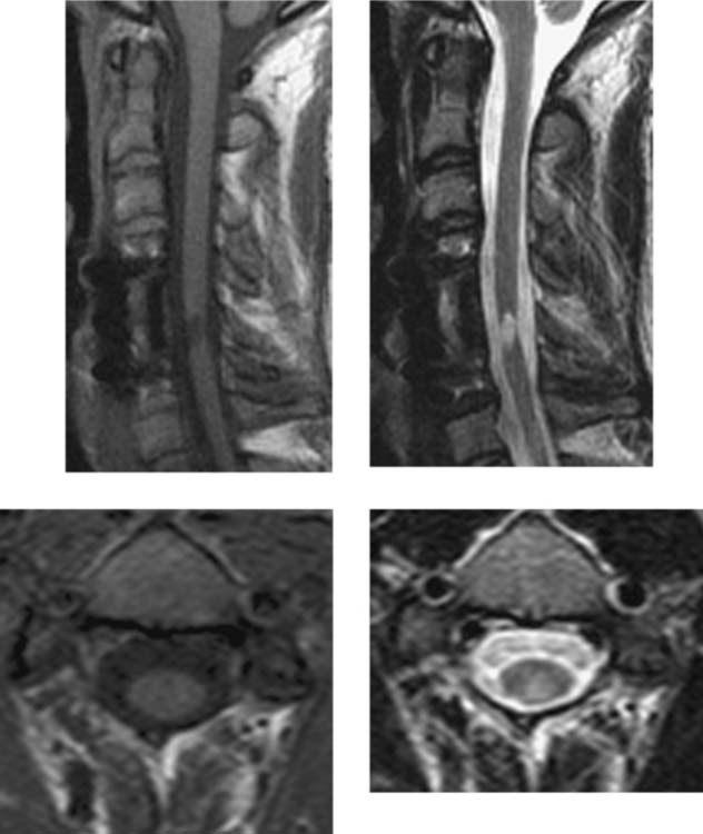
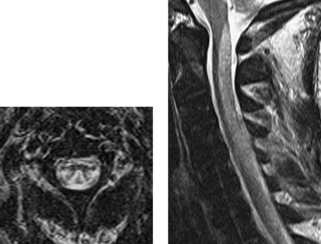
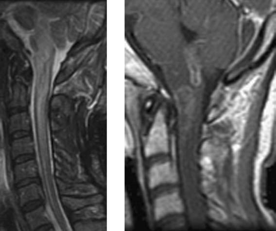
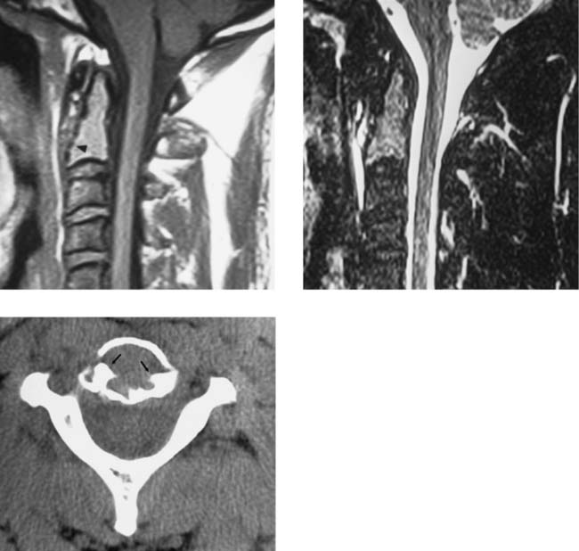
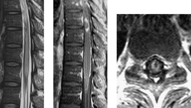
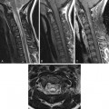
The patient has had an anterior fusion from C4 to C6. This was done following a motor vehicle accident to treat a C5 vertebral body fracture. The focal area of abnormal cord signal at C5–C6 is isointense to CSF on the T1W and T2W images, and there is associated mild cord atrophy at this level. These findings are consistent with a posttraumatic syrinx and cord tissue loss. Contiguous with this lesion and extending superiorly in the territory of the posterior columns is another region of abnormal signal that is isointense (to uninvolved cord) on the T1W and hyperintense on the T2W axial images (C3 level). This region extends to the cervicomedullary junction and is consistent with wallerian degeneration in the posterior columns.


Wallerian degeneration refers to antegrade degeneration of axons and their accompanying myelin sheaths and results from injury to the proximal portion of the axon or its cell body. The MR manifestations and temporal course of wallerian degeneration that occur above and below a spinal cord injury have been described by Quencer and colleagues in a study comparing MR and histologic results from formalin-fixed postmortem human cords (N = 24). MR images showed increased signal intensity in the posterior columns above the injury level, and in the lateral corticospinal tracts below the injury level, in all cases in which cord injury had occurred 7 or more weeks before death. Injuries occurring 8–12 days before death showed no abnormal signal above or below the level, although early wallerian degeneration was present histologically. Thus, in the injured spinal cord, wallerian degeneration is unlikely to be detected between 8 days and 7 weeks postinjury, yet should become apparent at time intervals greater than 7 weeks postinjury. Interestingly, the injured cords did not show the low signal intensity that has been observed on T2W images of cerebral white matter in vivo at 4–14 weeks following cerebral infarction. In the brain, wallerian degeneration produces hyperintense white matter signal at time intervals greater than 14 weeks postinjury.


Notes


Vertebral Body Avascular Necrosis


Unilateral Cord Infarction, Cervical


Comment


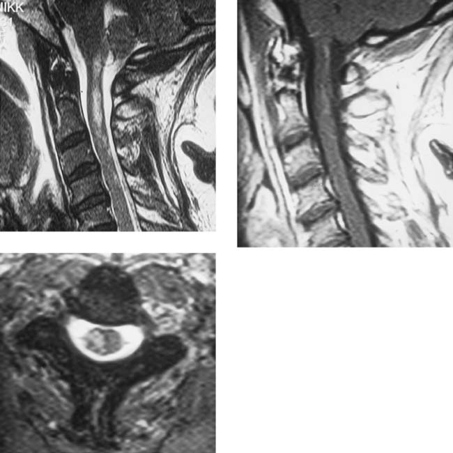
The distal right vertebral artery in this 43-year-old woman was embolized to control the loss of blood from a large posterior inferior communicating artery aneurysm that had ruptured during attempted embolization. Subsequently, she developed a right-sided, partial Brown-Séquard syndrome, affecting upper and lower extremities. The MR imaging study was obtained 5 days after embolization and shows hyperintensity on the right side of the cord at C1–C2 on the T2W sagittal and T2*W axial (C2 level) images. The axial image demonstrates the abrupt margin of the lesion at the midline of the cord and some sparing of the white matter of the posterior and lateral columns. Minimal enhancement is observed at C1–C2 on the postcontrast T1W image, without obvious cord enlargement. The findings are not typical of demyelinating disease, which should exhibit predominantly peripheral (white matter) rather than central (gray matter) involvement, or for intramedullary primary tumor (astrocytoma, ependymoma), which usually causes cord enlargement, more prominent enhancement than shown here, and cyst formation (≈25%–50% of cases).


The anterior spinal artery supplies the anterior two thirds of the cord parenchyma. It is derived from paired longitudinal vessels that fuse over most of their length by the second embryonic month. Unfused portions appear as duplications or fenestrations, most often seen in the cervical region. In the upper cervical spine, anterior medullary (or radiculomedullary) arteries from the vertebral arteries supply the anterior spinal artery. A plausible explanation for the findings in this case is that the patient has a duplicated cervical segment of the anterior spinal artery and that this segment is supplied by radiculomedullary branches of the right vertebral artery. Occlusion of the right vertebral artery caused an abrupt loss of blood to the right limb of the anterior spinal artery and ultimately resulted in a unilateral infarct and symptoms of a partial Brown-Séquard syndrome. A similar mechanism has been proposed to explain unilateral cord infarction in a patient with ipsilateral vertebral artery dissection and congenital afibrinogenemia (Laufs et al). Unilateral cervical cord infarctions can also occur with a single anterior spinal artery. The occluded vessel (or vessels) in this case is a sulcal artery that originates from the anterior spinal artery in the anterior median fissure and courses centrally and then to the right or left to supply central gray matter. Because adjacent sulcal arteries alternate their supply to the right and left sides of the cord (right, then left, then right for successive sulcal arteries) and because the single anterior spinal artery is supplied by both vertebral arteries, it is rare to have a unilateral infarct resulting from occlusion of a single vertebral artery.


The paired posterior spinal arteries supply the posterior third of the cord parenchyma, including the posterior columns, posterior horns, and posterolateral portion of the lateral columns. In the upper cervical spine these arteries are supplied by posterior medullary (or radiculopial) arteries, which originate primarily from the vertebral arteries. Because the posterior spinal arteries are part of the rich anastomotic pial network on the cord surface, it is unusual to have a posterior cord infarct. Examples of a single posterior medullary artery supplying both posterior spinal arteries have been reported, however, and in one case (Bergqvist et al), right vertebral artery dissection resulted in bilateral posterior spinal artery territory infarction and a posterior cord syndrome.


Notes


Acute Spinal Subdural Hematoma


Comment


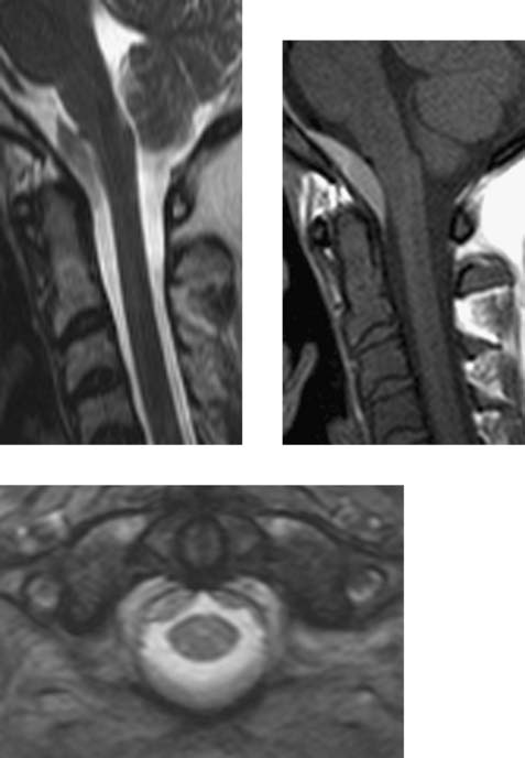
The lesion anterior to the cervicomedullary junction is hyperintense on the T1W image and hypointense on the T2W sagittal image. On the gradient-echo (GRE) T2*W axial image, the abnormal collection is deep to the dura (which is seen as a low-intensity arc encompassing the anterior half of the thecal sac) and has a scalloped appearance. Acute spinal subdural hematoma (ASSH) is relatively uncommon. It is most often detected in the thoracolumbar spine. Risk factors include trauma, coagulopathy, recent lumbar puncture, recent epidural anesthesia, and ruptured vascular malformation. ASSH has been observed in patients following minor trauma, such as sneezing or coughing. Frequently, presenting symptoms include acute motor and sensory impairment and loss of sphincter tone. Surgical decompression is the preferred treatment; however, conservative treatment is advocated if the presenting symptoms are either mild or resolving. Resorption occurs approximately 2 to 16 weeks after presentation. Distinguishing between an ASSH and the more common epidural hematoma is frequently difficult. Signs that favor a subdural hematoma include lack of “capping” by the epidural fat and lack of direct continuity of the collection with the adjacent vertebral body. Signal intensity of the collection is variable on both T1W and T2W images, although low signal is frequently seen on fast-spin-echo (FSE) T2W and GRE T2*W images. In the older study by Post et al, the distinction between a subdural and an epidural hematoma was easier on CT, and the authors concluded that CT and MR were complementary techniques.


Notes


Intraspinal Gas Collection


Comment


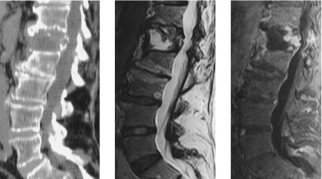
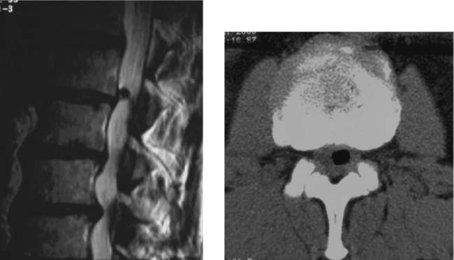
The left parasagittal T2W image shows focal low signal intensity posterior and superior to the L1–L2 disk space. This feature could represent gas, hemorrhage, calcification, or a vascular flow void. The hyperintense superior rim of the lesion is a susceptibility artifact. On the CT image, the lesion is markedly hypodense, consistent with a gas collection. Although the gas collection contacts the disk space, the presence or absence of a thin rim of “soft” disk material surrounding the gas cannot be determined. No “vacuum phenomenon” is present within the L1–L2 disk space. It is often assumed that a gas collection like the one shown here must be associated with a disk herniation, either extradural or, less commonly, intradural. Some authors, however, have proposed a few etiologies for symptoms of disk herniation when a gas collection is present. These include (1) free gas in the epidural space causing nerve root compression, (2) “gas bubbles” with a reactive peripheral fibrous capsule, and (3) herniation of the nucleus pulposus of an intervertebral disk that already has a vacuum phenomenon.


The vacuum phenomenon within an intervertebral space represents the accumulation of gas in preexisting diskal fissures or cavities in response to decreased intradiskal pressure caused by external forces applied to the spine or by hyperextension of the spine. Intervertebral gas collections reportedly increase with extension and decrease with flexion of the spine. They are composed of nitrogen (90–92%) combined primarily with oxygen and carbon dioxide. Intraspinal gas collections that appear to be causing nerve root compression have been “treated” by percutaneous aspiration with an 18-gauge spinal needle. Preliminary results indicate that symptoms can be alleviated for at least several months.


Notes


Radiation Myelopathy


Comment


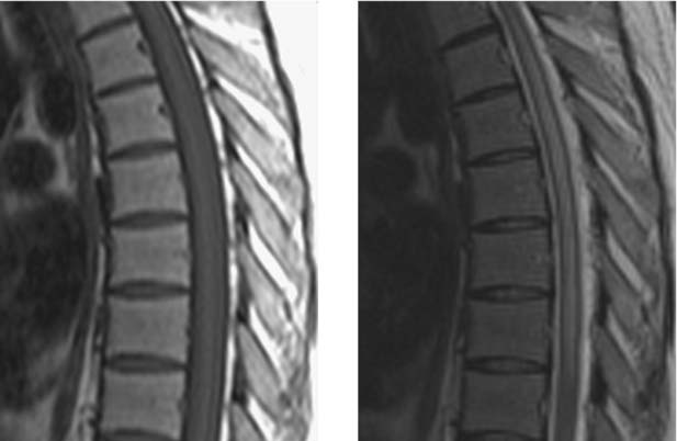
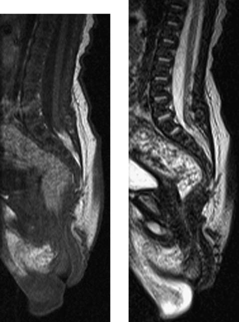
Imaging of the midthoracic region reveals mild, diffuse cord enlargement, and diffuse cord hyperintensity (edema) on the T2W image. Radiation myelopathy is a diagnosis of exclusion, and other etiologies, such as neoplastic disease (primary or metastatic) and infectious disease, must be ruled out before diagnosis. Diffuse abnormal hyperintensity of the vertebral bodies on the T1W images, though, is a key finding that can narrow the differential diagnosis. The latency period between completion of radiation therapy and onset of symptoms ranges from a few weeks to 12 years. Most cases, however, present between 6 months and 2 years after radiation treatment. A minimal dose of 50 Gy to the cord is usually required. Typical symptoms include ascending sensorimotor symptoms. These include Brown-Séquard syndrome, hemiparesis, bulbar palsy, and quadriparesis. Symptoms tend to be progressive and irreversible.


Based on MR imaging of patients who developed cervical myelopathy following radiotherapy for nasopharyngeal carcinoma, Wang et al noted a correlation between the time of MR imaging after the onset of symptoms and the MR findings. MR imaging performed less than 8 months after the onset of symptoms typically revealed hypointensity within the cord on T1W images and corresponding hyperintensity on T2W images. The signal intensity changes occurred with or without associated swelling of the cord and focal postcontrast enhancement. MR imaging performed more than 3 years after the onset of symptoms usually demonstrated cord atrophy without abnormal signal intensity.


Notes


Polyostotic Fibrous Dysplasia


Comment


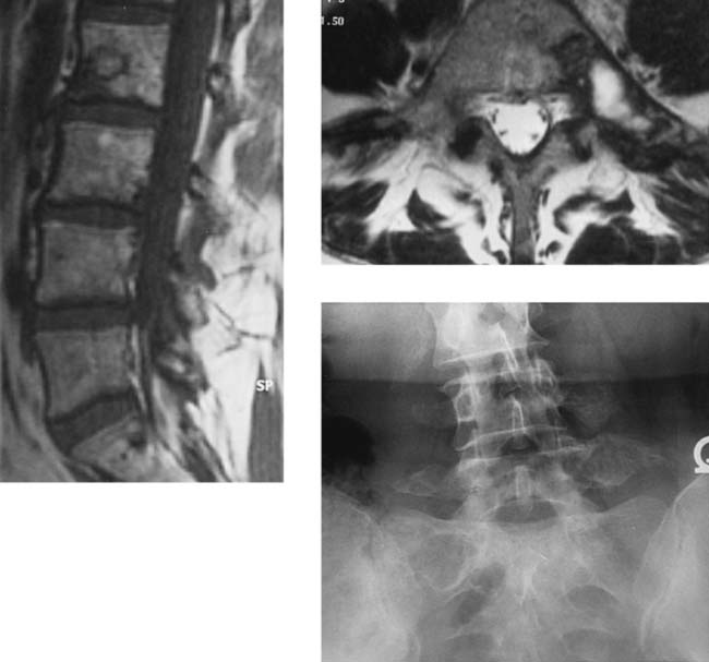
The sagittal T1W image shows an L2 body lesion with isointense center and hypointense rim, and a smaller S1 lesion. On the axial T2W image, a lesion involving the body, left pedicle, and transverse process of L5 has a hyperintense center and extensive hypointense periphery. The radiograph reveals expansile, trabeculated lesions of the left L5 and L4 transverse processes and a relatively lucent left L5 pedicle. The right side of L3 is expanded, with increased density, whereas the right sacral ala of S1 has a radiolucent lesion. Typically, MR signal intensity in fibrous dysplasia (FD) is variable and depends on the relative amounts of fibrous, cartilaginous, and (sometimes) hemorrhagic components. Lesions are isointense/hypointense on T1W images. On T2W images, fibrous regions are hypointense, whereas cartilaginous regions are hyperintense. In general, FD is more commonly monostotic (80–85%) than polyostotic (15–20%). The association of polyostotic involvement with cutaneous and/or endocrine (hyperthyroidism, Cushing syndrome, sexual precocity) manifestations is termed the McCune-Albright syndrome. It occurs in up to 50% of females with polyostotic FD. Malignant transformation of FD to sarcoma is uncommon (0.5%). In the spine, FD is more frequently polyostotic than monostotic, and the prevalence of polyostotic disease may be as high as 63%. The order of involvement is lumbar image thoracic image sacral image cervical (see Leet et al). Monostotic FD of the spine is exceedingly rare (< 30 cases), usually involves the vertebral body and adjacent pedicle, and shows no predilection for a particular spinal region. Most spinal FD lesions are asymptomatic and require no treatment. Many patients do have spinal pain, and a few pathologic compression fractures, with or without trauma, have been reported. Scoliosis is common in patients with polyostotic FD.


Notes


Subacute Combined Degeneration (SCD)


Listeria Myelitis/Rhombencephalitis


Retrosomatic Cleft


Comment


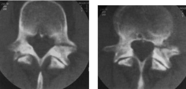
The CT images demonstrate a pedicular defect that has relatively smooth margins and no definite evidence of healing. There was no history of trauma. The cause of retrosomatic clefts is uncertain. Many authors believe that the defects represent congenital anomalies, while others argue that fractures are responsible. The lack of a history of trauma in many cases, the coexistence of other vertebral anomalies, and a published report describing cartilage obtained from the cleft support a congenital etiology. Retrosomatic cleft involves the pedicle and may result from anomalous ossification centers. The affected pedicle may be elongated, shortened, or thickened. It is most commonly found in women older than 30 years. Its location anterior to the transverse process differentiates it from spondylolysis, retroisthmic cleft, and spina bifida (all located posterior to the transverse process). Its pedicular location differentiates it from persistent neurocentral synchondrosis, which represents a failure of fusion of the three vertebral body ossification centers. Its coronal orientation, usual short length, and smooth margins differentiate it from pedicular hypoplasia and aplasia. Hypertrophic changes adjacent to the cleft may be present. It has been suggested that retrosomatic clefts at a single vertebral level are of no clinical significance unless there is associated disk degeneration. Retrosomatic clefts have been reported from T12 through L5. A few cases of bilateral clefts at one level, and a case of bilateral clefts at three consecutive levels (L3, L4, and L5) have been published.


Notes


Acute Calcific Prevertebral Tendinitis (Calcific Tendinitis of the Longus Colli Muscle)


Charcot-Marie-Tooth Disease (Hereditary Motor and Sensory Neuropathy Type I)


Comment


The images show bilateral enlargement of the spinal nerve roots and peripheral nerves in a 60-year-old man. At the L5 vertebral level, intradural nerve root enlargement (L5 roots laterally) and paraspinal nerve enlargement (L4 ventral rami, arrow) are evident. At the S1 level, the roots in the neural foramina are enlarged, as are the lumbosacral trunks (arrow points to left lumbosacral trunk, with adjacent vessels). The hereditary hypertrophic neuropathies include Charcot-Marie-Tooth disease type I (CMT I, HMSN type I), Dejerine-Sottas disease (HMSN type III), and Refsum disease (HMSN type IV). HMSNs are heterogeneous disorders characterized by chronic degeneration of peripheral nerves and roots, with subsequent muscle atrophy and sensory impairment in a distal distribution. Nerve enlargement may be the result of cycles of demyelination followed by repair and remyelination. CMT disease is usually inherited as an autosomal dominant trait, and onset is in late childhood or adolescence. In patients with CMT I, the peripheral nerves are characterized by variable enlargement and slowed conduction. MR findings differ from patient to patient, with variable combinations of nerve enlargement, hyperintensity on T2W images, and postcontrast enhancement in most patients and negative findings in others. In a study of 7 CMT patients, Cellerini et al found that hypertrophic nerves in CMT I tend to enhance, and correlations with biopsy results suggest that the enhancement is not related to inflammatory infiltrates but rather to disruption of the blood-nerve barrier from congenital or demyelinating processes. In patients with CMT II, peripheral nerves had normal size, electrical conduction, and appearance on MR imaging. The differential diagnosis for diffuse intradural nerve root enhancement and enlargement includes chronic inflammatory demyelinating polyneuropathy (CIDP), meningeal carcinomatosis, lymphoma, amyloidosis, sarcoidosis, neurofibromatosis type 1, and leprosy. Enhancement without enlargement may be seen with Guillain-Barré syndrome, CIDP, infective polyradiculopathies such as cytomegalovirus (CMV) polyradiculoneuritis, postsurgical arachnoiditis, and radiation therapy.


Notes


Expansive Open-Door Laminoplasty


Comment


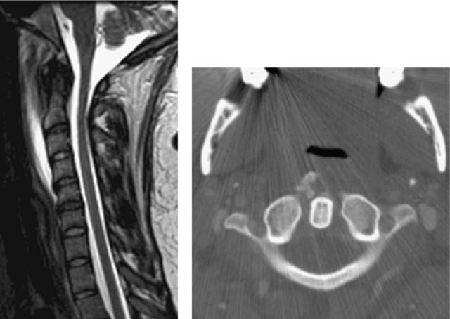
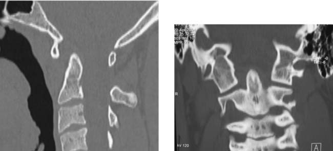
In the first patient, pretreatment T2W imaging reveals multilevel canal stenosis due to osteophytic ridging and thick hypointensity posterior to the vertebral bodies, suggesting some ossification of the posterior longitudinal ligament (OPLL). Comparison with CT images and plain radiographs (not shown here) should be done to confirm this diagnosis. Focal hyperintensity in the cord at C3–C4 on the pretreatment image is resolved on the posttreatment image, consistent with a decrease in edema. In the second patient, the axial CT image at C3 reveals changes in the usual appearance of the cervical spine. Both patients have undergone a modified version of a surgical procedure called expansive open-door laminoplasty, which was first described by Hirabayashi in 1977. This procedure has been used primarily to treat symptomatic, multilevel cervical canal stenosis of various etiologies. Typically the C3 to C7 vertebrae are altered. C2 is left intact because of the attachment of paraspinal muscles necessary for neck stability. Hence, there is a step-off in the spinolaminal line between the intact C2 and the altered C3 posterior elements on lateral radiographs. As shown on the CT image, the right side of the canal is “opened” by resecting most of the ipsilateral lamina. This results in an increase in the cross-sectional area of the canal. Opening of the canal is facilitated by burring through the outer cortex of the bone at the contralateral lamina-facet junction, thereby creating a “hinge.” The result is an “open door” side and a “hinge side” of the altered vertebrae. On the “open door” side, there is greater access to the neural foramina so that unilateral foraminotomies may be performed at the time of surgery to ameliorate ipsilateral radiculopathic symptoms. A rib allograft is used to keep the “door” open. Typically, these grafts are located at C3, C5, and C7. Optimal widening of the anteroposterior diameter of the spinal canal by expansive laminoplasty is considered to be over 4 mm.


Notes


Plasmacytoma


Comment


The sagittal T1W and STIR images demonstrate abnormal, increased signal intensity within the C2 body and odontoid process, and a prominent, irregular hypointense margin corresponding to bony cortex (arrowhead), confirmed on the CT scan. The central portion of the C2 vertebral lesion is relatively homogeneous. Note the internal projections (arrows) of the residual bone of C2 on the CT image. Cortical bone destruction is present on CT, yet spared compared with cancellous bone destruction. The posterior elements of C2 are also spared. The bone scan in this patient showed no increased uptake in C2, and no other bone lesions were identified. Serum protein electrophoresis demonstrated a monoclonal immunoglobulin G peak.


Solitary plasmacytoma is uncommon and occurs in approximately 5% of patients with plasma cell myeloma. By strict definition, the diagnosis requires histologic evidence of a monoclonal plasma cell infiltrate in one bone lesion, absence of other bone lesions, and lack of marrow plasmacytosis elsewhere. While local radiotherapy is effective for the primary tumor, multiple myeloma develops in most patients within a few years. This C2 lesion differs somewhat from the typical vertebral plasmacytoma, which is expansile and hypointense on T1W images. A finding on axial MR images that has been reported as characteristic of solitary vertebral plasmacytoma is the “mini-brain” pattern. In this pattern, curvilinear structures with low signal intensity on all imaging sequences extend partially through the vertebral body. These structures, which probably represent thick cortical struts resulting from compensatory hypertrophy of residual trabecular bone, resemble sulci seen in the brain. The pattern has not been described for other primary or metastatic bone lesions and is postulated to result from the less aggressive nature of plasmacytoma. The internal projections (arrows) of residual bone in this case are more blunted and less abundant than the struts that characterize the mini-brain appearance in published reports. Based on the C2 signal intensity on the sagittal MR images, one may initially consider vertebral hemangioma in the differential diagnosis; however, hemangiomas typically show a honeycomb pattern on CT rather than the lytic lesion seen here. The less common, aggressive form of hemangioma has predominantly angiomatous stroma and may appear as a lytic lesion with soft tissue density on CT. Aggressive hemangioma, however, is usually hypointense on T1W images.


Notes


Vestigial Tail


Retroisthmic Cleft


Comment


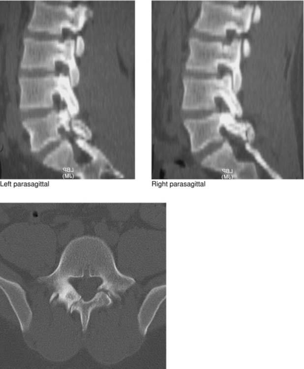
As illustrated in this case, the retroisthmic cleft is characterized by regular (or sometimes irregular) osseous margins, hypertrophic changes (small spurs) at the defect, and sclerosis and thickening of the contiguous neural arch. The defect is located in the right lamina posterior and inferior to the pars interarticularis. Parasagittal images, as shown here, are helpful in differentiating a retroisthmic defect, which is posterior to the lower facet joint, from a pars defect, which is anterior and superior to the joint. Oblique sagittal reconstructed images (not shown) along the axis of the involved lamina are also helpful in diagnosis. Note that the parasagittal images demonstrate a mild anterior subluxation (spondylolisthesis) of L5 on S1.


Of the 12 cases of retroisthmic cleft reported in the literature up to the year 2000, four cases had associated unilateral, contralateral spondylolysis. Thus, the frequency of contralateral spondylolysis was at least five times the prevalence (5% to 7%) of spondylolysis occurring in the general population. In all reported cases, retroisthmic clefts have been identified only in the L5 (9 of 12) and L4 (3 of 12) vertebrae. On the basis of these and other observations, as well as a single case demonstrating radiographic changes in a laminar defect over 6 years and increased activity in the region of the defect on bone scan, Wick and colleagues have argued that the cause of a retroisthmic defect is a stress fracture caused by chronic mechanical overload (of a weakened vertebral arch). This etiology, referred to as laminolysis, is analogous to mechanisms proposed for the development of the pars interarticularis cleft (spondylolysis) and the retrosomatic cleft (pediculolysis). Previous reports of retroisthmic and retrosomatic clefts considered them to be congenital anomalies. On MR imaging, both T1W and T2W images show the retroisthmic cleft as a line of decreased signal intensity with well-defined, sharp borders in a lamina.


Notes


Atlantoaxial Dislocation with Occipitalized Atlas


Intramedullary Abscess


Feb 14, 2016 | Posted by in MUSCULOSKELETAL IMAGING | Comments Off on Challenge

Full access? Get Clinical Tree

Get Clinical Tree app for offline access