Chance Fracture
Benjamin Y. Huang
CLINICAL HISTORY
8-year-old female involved in a head-on motor vehicle collision. The patient was a restrained rear-seat passenger. She currently complains of pain in her midback.
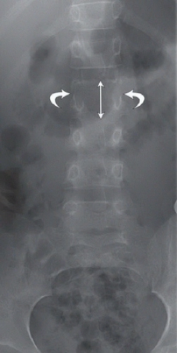 FIGURE 33A |
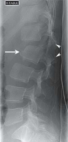 FIGURE 33B |
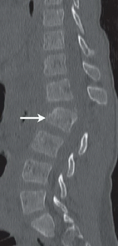 FIGURE 33C |
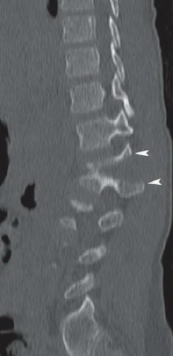 FIGURE 33D |
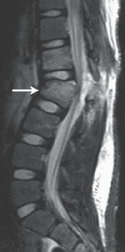 FIGURE 33E |
FINDINGS
Figure 33A: Anteroposterior (AP) radiograph of the lumbar spine demonstrates splitting of the bilateral L2 pedicles (curved arrows) as well as widening of the L1-L2 interspinous space. Figure 33B: Cross-table lateral lumbar spine radiograph demonstrates anterior wedging of the L2 vertebral body (arrow). There are markedly distracted, horizontally oriented fractures that extend through the pedicles and interarticular processes of L2 (arrowheads). There is also anterolisthesis of L1 on L2 with widening of the posterior disk space, causing focal kyphosis. Figures 33C and 33D: Sagittal reformatted CT images of the lumbar spine again demonstrate wedging of the L2 vertebral body (arrow
in Fig. 33C) and widening of the interspinous space. The parasagittal CT image (Fig. 33D) demonstrates the distracted fracture line extending through and vertically splitting the left posterior vertebral arch (arrowheads). Figure 33E: Midsagittal STIR image of the lumbar spine demonstrates anterior compression of the L2 vertebral body (arrow). There is edema within the vertebral body as well as in the L1 vertebra above. More posteriorly, there is disruption of the posterior longitudinal ligament at its attachment to the posterior-superior aspect of L2 and of ligamentum flavum. There is extensive edema in the paraspinous soft tissues.
in Fig. 33C) and widening of the interspinous space. The parasagittal CT image (Fig. 33D) demonstrates the distracted fracture line extending through and vertically splitting the left posterior vertebral arch (arrowheads). Figure 33E: Midsagittal STIR image of the lumbar spine demonstrates anterior compression of the L2 vertebral body (arrow). There is edema within the vertebral body as well as in the L1 vertebra above. More posteriorly, there is disruption of the posterior longitudinal ligament at its attachment to the posterior-superior aspect of L2 and of ligamentum flavum. There is extensive edema in the paraspinous soft tissues.
Related posts:


Stay updated, free articles. Join our Telegram channel

Full access? Get Clinical Tree


