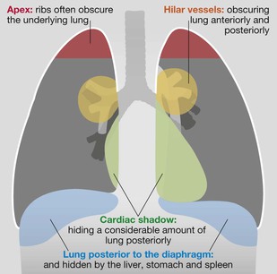
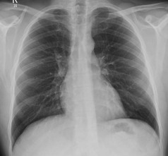
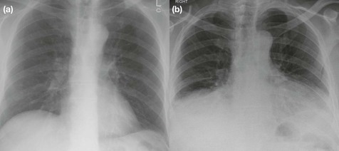
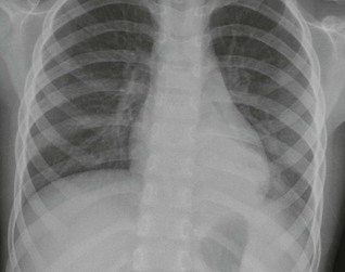
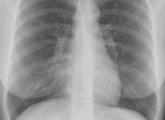
The tricky hidden areas. The four areas where small (and large) lung lesions are overlooked. Normal lung markings are solely due to vessels and to nothing else. The normal bronchial walls, normal interstitium, and normal lymphatics are not visible. There is a complete absence of any lung markings in the lung immediately adjacent to each costophrenic angle. In this peripheral part of the lung the normal vessels are simply too small to be visualised. If this area shows detectable markings, then interstitial disease, or oedema, is likely1. The hilum is the site at which the bronchi and vessels enter or leave the lung. Most normal hearts have a cardiothoracic ratio (CTR) of less than 50% when assessed on a PA chest radiograph obtained in full inspiration2. The two fissures in the right lung divide the lung into three lobes. The single fissure in the left lung divides the lung into two lobes. The oblique fissure on each side is propeller shaped and consequently we only, and very occasionally, see a part of a fissure on a normal lateral CXR. On the other hand, we will see most of the horizontal fissure on the lateral CXR because it is straight. We will often see this fissure on the frontal CXR for the same reason. Four steps underpin accurate analysis. 4. Now you can assess the radiograph in an organised and systematic manner. As follows: □ Is the heart enlarged? □ Are both domes of the diaphragm clearly seen and well defined? □ Are both heart borders clearly seen and well defined? □ Are the hila normal … position, size, density? □ Check the tricky hidden areas of the lung: both apices, behind the heart shadow, around each hilum, below the diaphragm (see p. 308). □ Finally, ask yourself once again: Have I addressed this patient’s particular clinical problem? Is there an adequate inspiration? PA CXRs of the same patient: (a) is a good inspiration; (b) is a very poor inspiration. In (b) the heart appears enlarged and the lung bases hazy with crowding of vessels. These appearances are bogus; they are due to the extremely poor inspiration. CXR (a) is normal. Are both domes of the diaphragm clearly seen and well defined? Both domes of the diaphragm will be well defined if the adjacent lung is normal. In this patient, presenting with chest pain and fever, much of the left dome is obscured. This is due to pus in the adjacent alveoli. Diagnosis: left lower lobe pneumonia. Are both heart borders clearly seen and well defined? Both heart borders will be well defined if the adjacent lung is normal. In this patient with a cough and fever the right heart border is obscured because the air in the adjacent lung has been replaced by pus. Diagnosis: middle lobe pneumonia. Four questions underpin accurate analysis: 1. Are the vertebral bodies becoming blacker from above downwards? 3. Is there any abrupt change in density across the heart shadow? 4.
Chest
Normal anatomy
Frontal CXR—the lungs
Frontal CXR—lung markings
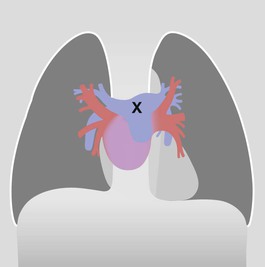
Frontal CXR—hila
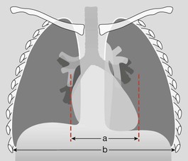
Frontal CXR—cardiothoracic ratio
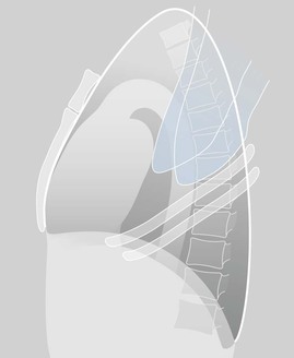
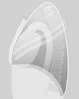
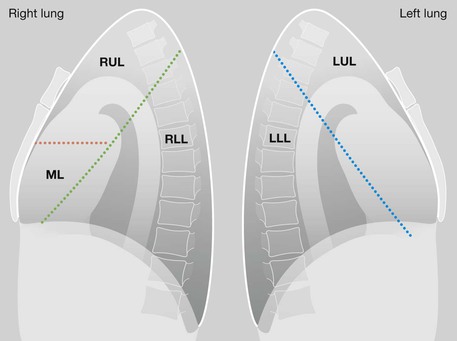
Lateral CXR—the lobes of the lung
Lateral CXR—skeletal shadows
Analysis: the checklists
The frontal CXR
In an adult, the cardiothoracic ratio (CTR) should be < 50% on a PA CXR (see p. 309).
If part of a dome is obscured, suspect pathology in the adjacent lower lobe.
If not, there is a high probability of pathology in the immediately adjacent lung.
The lateral CXR
If not (ie they are becoming whiter or greyer) then suspect disease in a lower lobe or in a pleural space.
“An abrupt change in density” is likely to be a lung abnormality.
Related posts:
![]()
Stay updated, free articles. Join our Telegram channel

Full access? Get Clinical Tree


Chest
18