Chapter Outline
Imaging Anatomy, 1
Gross Lung Anatomy, 1
Parenchymal Anatomy, 4
Pulmonary Function, 5
Mediastinum, 5
Imaging Protocols, 5
Infection, 6
Acquired Immunodeficiency Syndrome, 18
Neoplasm, 20
General, 20
Bronchogenic Carcinoma, 21
Tumor Staging, 22
Specific Lung Tumors, 25
Lung Metastases From Other Primary Lesions, 27
Chronic Lung Disease, 27
Idiopathic Diseases, 27
Lymphoproliferative Disorders, 31
Collagen Vascular Diseases, 32
Vasculitis and Granulomatoses, 33
Other Chronic Disorders, 34
Inhalational Lung Disease, 35
Pneumoconiosis, 35
Antigen-Antibody–Mediated Lung Disease, 38
Toxin-Induced Interstitial Pneumonitis/Fibrosis, 39
Airway Disease, 39
Chronic Bronchial Disease, 41
Lung Injury, 44
Postoperative Chest, 47
Pulmonary Vasculature, 49
Pulmonary Arterial Hypertension, 49
Pulmonary Edema, 50
Pulmonary Embolism, 51
Vasculitis, 52
Venous Abnormalities, 52
Pleura, 53
Mediastinum, 57
General, 57
Anterior Mediastinal Tumors, 57
Middle Mediastinal Tumors, 60
Posterior Mediastinal Tumors, 61
Other Mediastinal Disorders, 62
Differential Diagnosis, 62
General, 62
Atelectasis, 63
Consolidation, 64
Pulmonary Masses, 66
Cystic and Cavitary Lesions, 68
Interstitial Lung Disease, 70
Abnormal Density, 72
Tracheobronchial Lesions, 73
Pleural Disease, 74
Mediastinum, 74
Imaging Anatomy
Gross Lung Anatomy
Segmental Anatomy ( Figs. 1.1 – 1.2 )


Right Lung
| Upper lobe | Apical | B1 |
| Anterior | B2 | |
| Posterior | B3 | |
| Middle lobe | Lateral | B4 |
| Medial | B5 | |
| Lower lobe | Superior | B6 |
| Medial basal | B7 | |
| Anterior basal | B8 | |
| Lateral basal | B9 | |
| Posterior basal | B10 |
Left Lung
| Upper lobe | ||
| Upper | Apicoposterior | B1, B3 |
| Anterior | B2 | |
| Lingula | Superior | B4 |
| Inferior | B5 | |
| Lower lobe | Superior | B6 |
| Medial basal | B7 | |
| Anterior basal | B8 | |
| Lateral basal | B9 | |
| Posterior basal | B10 |
Segmental Computed Tomography (CT) Anatomy ( Fig. 1.3 )

Bronchial CT Anatomy ( Fig. 1.4 )

Plain Radiograph Anatomic Landmarks ( Figs. 1.5–1.9 )


Thoracic Inlet
The thoracic inlet represents the junction between structures at the base of the neck and those of the thorax. It parallels the first rib and is higher posteriorly than anteriorly.
Lines
- •
Anterior junction line: 2-mm linear line that projects over the trachea. Represents the approximation of the visceral and parietal pleura of the right and left lungs anterior to the mediastinum (composed of four layers of pleura)
- •
Posterior junction line (four layers of pleura): extends above clavicles and can often be seen on a frontal radiograph as a vertical line traversing the tracheal air column
- •
Posterior tracheal stripe (normally measures <4 mm in diameter): thickening or presence of a focal opacity in the region of the posterior tracheal stripe should raise the possibility of esophageal carcinoma
- •
Azygoesophageal line: interface between RLL air and mediastinum
- •
Left paraspinal line: extends from aortic arch to diaphragm
- •
Right paraspinal line
Paratracheal Stripe
- •
Abnormal if >4 mm
- •
Never extends below right bronchus
Fissures
The normal major fissures are seldom seen on a posteroanterior radiograph.
The top of the left lower lobe (LLL) is usually higher than the top of the right lower lobe (RLL).
- •
Minor (horizontal) fissure
- •
Major (oblique) fissure
- •
Azygos fissure
- •
Other fissures
Superior accessory fissure
Inferior accessory fissure
Left minor fissure
Pulmonary Ligament
- •
Consists of a double layer of pleura that connects the medial aspect of the lower lobe (LL) to the adjacent mediastinum and diaphragm
- •
Not seen on posteroanterior or lateral chest radiographs (CXRs)
- •
Determines the shape of the collapsed LL in patients with atelectasis and the shape of the collapsed lung in patients with pneumothorax
Trachea
- •
The trachea is a midline structure
- •
The aorta commonly causes a smooth indentation on the left side
- •
The trachea measures 10–12 cm in length
- •
16–20 U-shaped cartilage rings on its lateral and anterior aspects
- •
Calcification of the cartilage rings is a common normal finding in patients older than 40 years, particularly women, but it is seldom evident on radiographs
- •
Divides into the left and right main bronchi at the carina (approximately at the level of the fifth thoracic vertebra)
Upper Lobe (Ul) Bronchi ( Figs. 1.10 – 1.11 )
The right main bronchus divides into the RUL bronchus and the bronchus intermedius.
- •
RUL bronchus is always higher than LUL bronchus on lateral view
- •
Posterior wall of bronchus intermedius (right) is normally less than 2 mm thick and bifurcates into middle and LLs bronchi
- •
Tracheal bronchus (bronchus suis): 0.1% of population, arises from right wall of trachea (left much less common), supplies apical segment or occasionally entire RUL
- •
The left main bronchus is approximately 5 cm in length and divides into the LUL and LLL bronchi.
- •
Accessory cardiac bronchus: 0.1% of population, extends inferomedially from medial wall of bronchus intermedius or RLL bronchus toward mediastinum; may be blind ending


Parenchymal Anatomy
Acinus
- •
Includes all structures distal to one terminal bronchiole. The terminal bronchiole is the last purely air-conducting structure.
- •
Acinus measures 7 mm
- •
Acinus contains about 400 alveoli
Secondary Pulmonary Lobule
- •
Smallest anatomic unit of the lung visible on high-resolution CT (HRCT)
- •
Polygonal structure bounded by interlobular septa, 1.5–2 cm in diameter
- •
Three to five acini per secondary lobule
- •
Supplied by several terminal bronchioles
Epithelium
The alveolar epithelium is made up of two cell types:
- •
Type 1 pneumocytes
- •
Type 2 pneumocytes: produce surfactant, have phagocytic ability, and regenerate
- •
High-Resolution Computed Tomography (HRCT) ( Fig. 1.12 )

Technique
- •
1–1.5-mm thin collimation
- •
High spatial frequency reconstruction
This helps to improve spatial resolution, thereby improving the ability to detect subtle abnormalities—thick interlobular septa, cyst walls, small nodules, ground glass opacities and bronchiectasis.
- •
Optional
Increase in kVp or mA (140 kVp, 170 mA)
Targeted image reconstruction (one lung rather than both to increase spatial resolution)
HRCT Anatomy
The basic anatomic unit of pulmonary structure and function visible by HRCT is the secondary pulmonary lobule:
- •
Polyhedral 1.5-cm structure surrounded by connective tissue (interlobular septa) and made up of 5–15 pulmonary acini, which contain the alveoli for gas exchange
- •
Central artery and bronchiole
- •
Peripheral pulmonary veins and lymphatics in septum
Dominant high-resolution pattern:
- •
Reticular
- •
Nodular
- •
High attenuation (ground glass, consolidation)
- •
Low attenuation (emphysema, cystic)
Questions:
- •
Location within the secondary lobule
- •
Upper versus lower zone or a central versus peripheral predominance
- •
Presence of additional findings (pleural fluid, lymphadenopathy, traction bronchiectasis)
This protocol produces high-definition images of the lung alveoli, airways, interstitium, and pulmonary vasculature. Air trapping is identified on expiratory images.
Pulmonary Function ( Fig. 1.13 )

Lung Volumes, Capacities, and Flow Rates
- •
Tidal volume (TV): normal respiratory cycle
- •
Vital capacity (VC): amount of air that can be expired with force after maximal inspiration
- •
Functional residual capacity (FRC): volume remaining in lung after quiet expiration
- •
Total lung capacity (TLC): volume contained in lung at maximum inspiration
- •
Forced expiratory volume (FEV 1 ): amount of air expired in 1 second
Mediastinum ( Fig. 1.14 )
- •
Superior mediastinum: plane above aortic arch; thoracic inlet structures
- •
Anterior mediastinum: contains thymus, lymph nodes, mesenchymal tissue; some classifications include the heart and fat
- •
Middle mediastinum: contains heart, major vessels, trachea and main bronchi, lymph nodes, phrenic nerve, and left recurrent laryngeal nerve
- •
Posterior mediastinum: starts at anterior margin of vertebral bodies; contains descending thoracic aorta, esophagus, thoracic duct, azygos and hemiazygos veins, lymph nodes, autonomic nerves, paravertebral areas, and fat

Imaging Protocols
Standard Chest CT Protocol
Supine position. Scan in suspended inspiration at total lung capacity. Scan setup:
- •
5 × 5-mm sections from apex of the lungs to the adrenals
- •
Six 1.25-mm high-resolution cuts throughout lung at 2.5-cm intervals
- •
1-mm reconstructions through pulmonary nodules
- •
Number of different combinations of pitch and section thickness
In interstitial lung disease the six cuts are repeated with the patient in the prone position. Reconstruction is done with a high-resolution bone algorithm.
Use of IV contrast medium:
- •
Evaluation of vascular structures, arteriovenous malformation, aortic dissection
- •
Evaluation of mediastinal tumors, enlarged lymph nodes
- •
Hilar masses
- •
Neck masses
Pulmonary Embolism (PE) CT Protocol
- •
Patient in supine position
- •
Scan range: adrenals to lung apex
- •
Injection of 140 mL of nonionic iodinated contrast at 3 mL/second, with delay of 25–30 seconds. Scanning is performed with suspended respiration.
- •
Scans are retrospectively reconstructed from the dome of the diaphragm as 2.5-mm-thick slices with 1-mm spacing.
Diagnostic Radiology Report (American College of Radiology [ACR])
An authenticated written interpretation should be performed on all radiologic procedures. The report should include:
- 1.
Name of patient and other identifier (e.g., birth date, Social Security number, or hospital or office identification number)
- 2.
Name of the referring physician to provide more accurate routing of the report to one or more locations specified by the referring physician (e.g., hospital, office, clinic)
- 3.
History
- 4.
Name or type of examination
- 5.
Dates of the examination and transcription
- 6.
Time of the examination (for ICU/CCU patients) to identify multiple examinations (e.g., chest) that may be performed on a single day
- 7.
Body of the report:
- •
Procedures and materials
Include in the report a description of the procedures performed and any contrast media (agent, concentration, volume, and reaction, if any), medications, catheters, and devices.
- •
Findings
Use precise anatomic and radiologic terminology to accurately describe findings.
- •
Limitations
Where appropriate, identify factors that can limit the sensitivity and specificity of the examination. Such factors might include technical factors, patient anatomy, limitations of the technique, incomplete bowel preparation, and wrist examination for carpal scaphoid.
- •
Clinical issues
The report should address or answer any pertinent clinical issues raised in the request for the imaging examination. For example, to rule out pneumothorax state: “There is no evidence of pneumothorax.” To rule out fracture state: “There is no evidence of fracture.” It is not advisable to use such universal disclaimers as “The mammography examination does not exclude the possibility of cancer.”
- •
Comparative data
Comparisons with previous examinations and reports when possible are a part of the radiologic consultation and report and optionally may be part of the “impression” section.
- •
- 8.
Impression (conclusion or diagnosis):
- •
Each examination should contain an “impression” section.
- •
Give a precise diagnosis whenever possible.
- •
Give a differential diagnosis when appropriate.
- •
Recommend, only when appropriate, follow-up and additional diagnostic radiologic studies to clarify or confirm the impression.
In normal CXR section the only structures visible in normal lungs are the fissures and the pulmonary vessels.
Lung parenchymal abnormalities are divided into five basic patterns:
- 1.
Mass
- 2.
Consolidative
- 3.
Interstitial
- 4.
Vascular
- 5.
Airway
- 1.
- •
Infection
General
Pathogens
Bacterial pneumonia
- •
Streptococcus pneumoniae (pneumococcus)
- •
Staphylococcus
- •
Pseudomonas
- •
Klebsiella
- •
Nocardia
- •
Chlamydia
- •
Neisseria meningitides
- •
Haemophilus influenzae
- •
Anaerobes
- •
Legionella
- •
Mycoplasma pneumoniae
- •
Actinomyces israelii
- •
Mycobacterium tuberculosis
- •
Viral pneumonia (25% of community-acquired pneumonias)
- •
Influenza
- •
Varicella, herpes zoster
- •
Rubeola
- •
Cytomegalovirus
- •
Coxsackievirus, parainfluenza virus, adenovirus, respiratory syncytial virus (RSV)
- •
Fungal pneumonia
- •
Histoplasmosis
- •
Coccidioidomycosis
- •
Blastomycosis
- •
Aspergillosis
- •
Cryptococcosis
- •
Candidiasis
- •
Zygomycoses
- •
Parasitic pneumonias
- •
Pneumocystis jiroveci Frenkel 1999 (formerly Pneumocystis carinii )
- •
Toxoplasma gondii
- •
Acquisition of Pneumonia
Community-acquired pneumonia
- •
S. pneumoniae , Haemophilus
- •
Mycoplasma
- •
Hospital-acquired pneumonia (incidence 1%, mortality 35%): nosocomial infection
- •
Gram-negative bacteria: Pseudomonas , Proteus , Escherichia coli , Enterobacter , Klebsiella
- •
Methicillin-resistant Staphylococcus aureus (MRSA)
- •
Vancomycin-resistant enterococcus (VRE)
- •
Pneumonia in immunosuppressed patients
- •
Bacterial pneumonia (gram negative) still most common
- •
Tuberculosis
- •
Fungal
- •
Pneumocystis pneumonia (PCP)
- •
Endemic pneumonias
- •
Fungal: histoplasmosis, coccidioidomycosis, blastomycosis
- •
Viral
- •
Aspiration-associated pneumonia (important)
Risk Factors
The radiographic appearance of pulmonary infections is variable depending on the pathogen, underlying lung disease, risk factors, and previous or partial treatment.
| Risk Factor | Common Pathogens |
|---|---|
| Alcoholism | Gram-negative bacteria, Streptococcus pneumoniae , Mycobacterium tuberculosis , aspiration (mouth flora) |
| Old age | S. pneumoniae , Staphylococcus aureus , aspiration |
| Aspiration | Mouth flora (anaerobes) |
| Cystic fibrosis | Pseudomonas , S. aureus , Aspergillus |
| Chronic bronchitis | S. pneumoniae , Haemophilus influenzae |
Other risk factors for developing pneumonia:
- •
Bronchiectasis
- •
Coma, anesthesia, seizures (aspiration)
- •
Tracheotomy
- •
Antibiotic treatment
- •
Immunosuppression (renal failure, diabetes, cancer, steroids, AIDS)
- •
Chronic furunculosis ( Staphylococcus )
- •
Radiographic Spectrum of Pulmonary Infections


Complications of Pneumonia
- •
Parapneumonic effusion
Stage 1: exudation: free flowing
Stage 2: fibropurulent: loculated
Stage 3: organization, erosion into lung or chest wall
- •
Empyema
- •
Bronchopleural fistula (BPF; fistula between bronchus and pleural space) with eroding pleural-based fluid collections
- •
Bronchiectasis
- •
Pulmonary fibrosis, especially after necrotizing pneumonia or acute respiratory distress syndrome (ARDS)
- •
Adenopathy
Resolution of Pneumonia
- •
80%–90% of cases resolve within 4 weeks.
- •
5%–10% resolve within 4–8 weeks (usually in older or diabetic patients). Subsequent radiographs should always show interval improvement compared with the previous radiographs.
- •
Nonclearance
Antibiotic resistance
Consider other pathogen (e.g., M. tuberculosis )
Recurrent infection
Obstruction pneumonitis due to tumor
Bacterial Infections
General
Common Pathogens
- •
S. pneumoniae , 50% (40–60 years)
- •
Mycoplasma , 30%
- •
Anaerobes, 10%
- •
Gram-negative bacteria, 5%
- •
Staphylococcus , 5%
- •
Haemophilus , 3% (especially in infants and patients with chronic obstructive pulmonary disease [COPD])
Clinical Findings
Pneumonic syndrome
- •
Fever
- •
Cough
- •
Pleuritic pain
- •
Sputum
- •
Ancillary findings
- •
Headache, arthralgia, myalgia
- •
Diarrhea
- •
Hemoptysis
- •
Streptococcal Pneumonia
Radiographic Features
- •
Lobar or segmental pneumonia pattern
- •
Bronchopneumonia pattern
- •
Round pneumonia (in children)
Staphylococcal Pneumonia ( Fig. 1.15 )

Radiographic Features
- •
Bronchopneumonia pattern
- •
Bilateral, >60%
- •
Abscess cavities, 25%–75%
- •
Pleural effusion, empyema, 50%
- •
Pneumatoceles, 50% (check valve obstruction), particularly in children
- •
Central lines
- •
Signs of endocarditis
Pseudomonas Pneumonia
Typical Clinical Setting
- •
Hospital-acquired infection
- •
Ventilated patient
- •
Reduced host resistance
- •
Patients with cystic fibrosis
Radiographic Features
Three presentations:
- •
Extensive bilateral parenchymal consolidation (predilection for both LLs)
- •
Abscess formation
- •
Diffuse nodular disease (bacteremia with hematogenous spread; rare)
Legionnaires Disease
Severe pulmonary infection caused by Legionella pneumophila; 35% of patients require ventilation, 20% mortality. Most infections are community acquired. Patients have hyponatremia. Seroconversion for diagnosis takes 2 weeks.
Radiographic Features
Common features
- •
Initial presentation of peripheral patchy consolidation
- •
Bilateral severe disease
- •
Rapidly progressive
- •
Pleural effusions, <50%
- •
LL predilection
- •
Uncommon features
- •
Abscess formation
- •
Lymph node enlargement
- •
Haemophilus Pneumonia
Caused by H. influenzae . Occurs most commonly in children, immunocompromised adults, or patients with COPD. Often there is concomitant meningitis, epiglottitis, and bronchitis.
Radiographic Features
- •
Bronchopneumonia pattern
- •
LL predilection, often diffuse
- •
Empyema
Mycoplasma Pneumonia
Most common nonbacterial pneumonia (atypical pneumonia). Mild course. Age 5–20 years. Positive for cold agglutinins, 60%.
Radiographic Features
- •
Reticular pattern
- •
LL predominance, often diffuse
- •
Consolidation, 50%
Complications
- •
Autoimmune hemolytic anemia
- •
Erythema nodosum, erythema multiforme
- •
Stevens-Johnson syndrome
- •
Meningoencephalitis
Klebsiella (Friedländer) Pneumonia
Gram-negative organism. Often in debilitated patients and/or alcoholics.
Radiographic Features
- •
Consolidation appears similar to that of infection with S. pneumoniae
- •
Lobar expansion
- •
Cavitation, 30%–50%, typically multiple
- •
Massive necrosis (pulmonary gangrene)
- •
Pleural effusion uncommon
Tuberculosis (TB) ( Fig. 1.16 )

Transmitted by inhalation of infected droplets of M. tuberculosis or M. bovis . TB acquisition usually requires constant or repeated contact with sputum-positive patients because the tubercle does not easily grow in the immunocompetent human host. Target population includes:
- •
Patients of low socioeconomic status (homeless)
- •
Alcoholics
- •
Immigrants: from Mexico, Philippines, Indochina, Haiti
- •
Elderly patients
- •
AIDS patients
- •
Prisoners
Primary Infection ( Fig. 1.17 )
Usually heals without complications. Sequence of events includes:
- •
Pulmonary consolidation (1–7 cm); cavitation is rare; LL (60%) > UL
- •
Caseous necrosis 2–10 weeks after infection
- •
Lymphadenopathy (hilar and paratracheal), 95%
- •
Pleural effusion, 10%
- •
Spread of a primary focus occurs primarily in children or immunosuppressed patients.

Secondary Infection ( Fig. 1.18 )

Active disease in adults most commonly represents reactivation of a primary focus. However, primary disease is now also common in adults in developed countries because there is no exposure in childhood. Distribution is as follows:
- •
Typically limited to apical and posterior segments of ULs or superior segments of LLs (because of high P o 2 ?)
- •
Rarely in anterior segments of ULs (in contradistinction to histoplasmosis)
Radiographic Features
- •
Exudative TB
Patchy or confluent air space disease
Adenopathy uncommon
- •
Fibrocalcific TB
Sharply circumscribed linear densities radiating to hilum
- •
Cavitation, 40%
Complications ( Fig. 1.19 )
- •
Miliary TB may occur after primary or secondary hematogenous spread.
- •
Bronchogenic spread occurs after communication of the necrotic area with a bronchus; it produces an acinar pattern (irregular nodules approximately 5 mm in diameter).
- •
Tuberculoma (1–7 cm): nodule during primary or secondary TB; may contain calcification
- •
Effusions are often loculated.
- •
Bronchopleural fistula
- •
Pneumothorax
| Primary TB | Reinfection TB | |
|---|---|---|
| Location | Usually bases | Upper lobes, superior segment |
| Lower lobes | ||
| Appearance | Focal | Patchy |
| Cavitation | No | Frequent |
| Adenopathy as only finding | Common | No |
| Effusion | Common | Uncommon |
| Miliary pattern | Yes | Yes |

Nontuberculous Mycobacterial (NTMB) Infections
The two most common NTMB pathogens are M. avium-intracellulare and M. kansasii (less common: M. xenopi , M. chelonei , M. gordonae, M. fortuitum , “fast grower”). Unlike TB, NTMB infections are not acquired by human-human transmission but are a direct infection from soil or water. There is also no pattern of primary disease or reactivation: the infection is primary, although some infections may become chronic. The infection often occurs in elderly patients with COPD, older women in good health, and AIDS patients.
Radiographic Features
- •
NTMB infections may be indistinguishable from classic TB.
- •
Atypical features such as bronchiectasis and bronchial wall thickening are common.
- •
Nodules are common in older women.
COMPUTED TOMOGRAPHY FINDINGS
Findings
TB (%)
MAI Infection (%)
Nodules <1 cm
80
95
Nodules 1–3 cm
40
30
Mass >3 cm
10
10
Consolidation
50
50
Cavity
30
30
Bronchiectasis
30
95
Bronchial wall thickening
40
95
Septal thickening
50
15
Emphysema
20
20
Calcified granuloma
15
5
MAI , Mycobacterium avium-intracellulare; TB , tuberculosis.
Nocardia Pneumonia
Caused by Nocardia asteroides , worldwide distribution. Common opportunistic invader in:
- •
Lymphoma
- •
Steroid therapy; especially transplant patients
- •
Pulmonary alveolar proteinosis (common)
Radiographic Features
- •
Focal consolidation (more common)
- •
Cavitation
- •
Irregular nodules
Actinomycosis
Actinomycosis is caused by Actinomyces israelii, a gram-positive normal saprophyte in the oral cavity. Pulmonary disease develops from aspiration of the organism (poor dentition) or from direct penetration into the thorax.
Radiographic Features
- •
Focal consolidation > cavitating mass
- •
Lymphadenopathy uncommon
- •
Extension into the chest wall and pleural thickening is less common today but still occurs and is an important differential feature.
Pulmonary Abscess
The spectrum of anaerobic pulmonary infections includes:
- •
Abscess: single or multiple cavities >2 cm, usually with AFL
- •
Necrotizing pneumonia: analogous to abscess but more diffuse and cavities <2 cm
- •
Empyema: suppurative infection of the pleural space, most commonly as a result of pneumonia
Predisposing Conditions
- •
Aspiration (e.g., alcoholism, neurologic disease, coma)
- •
Intubation
- •
Bronchiectasis, bronchial obstruction
Treatment
- •
Antibiotics, postural drainage
- •
Percutaneous drainage of empyema
- •
Drainage/resection of lung abscess only if medical therapy fails
Sickle Cell Anemia
- •
Patients with sickle cell disease are at increased risk of pneumonia and infarction. These entities are difficult to differentiate, and hence are called acute chest syndrome .
- •
Pneumonias were originally due to pneumococci but now are due to viruses or Mycoplasma. Differential diagnosis includes atelectasis and infarct.
- •
Infarcts are more frequent in adults than in children. Rare in children younger than 12 years.
- •
Consolidation is seen on CXRs; resolves more slowly than in the general population and tends to recur.
Viral Pneumonia
General
Classification
DNA viruses
Unenveloped
- •
Parvoviruses
- •
Papovaviruses
- •
Adenoviruses
- •
Hepatitis viruses (hepatitis B)
- •
Enveloped
- •
Herpesviruses (herpes simplex virus [HSV], Epstein-Barr virus [EBV], varicella-zoster virus [VZV], cytomegalovirus [CMV])
- •
Poxviruses (variola virus, molluscum contagiosum virus)
- •
RNA viruses
Unenveloped
- •
Picornaviruses (hepatitis A virus, coxsackievirus)
- •
Caliciviruses
- •
Reoviruses
- •
Enveloped
- •
Retroviruses (HIV)
- •
Arenaviruses
- •
Coronaviruses
- •
Togaviruses
- •
Bunyaviruses
- •
Orthomyxoviruses (influenza virus)
- •
Paramyxoviruses (mumps virus, measles virus, RSV, parainfluenza virus)
- •
Occurrence
Immunocompetent hosts
Influenza
Hantavirus
EBV
Adenovirus
Immunocompromised hosts
HSV
VZ
CMV
Measles virus
Adenovirus
Spectrum of Disease
- •
Acute interstitial pneumonia (AIP): diffuse or patchy interstitial pattern, thickening of bronchi, thickened interlobar septa
- •
Lobular inflammatory reaction: multiple nodular opacities 5–6 mm (varicella; late calcification)
- •
Hemorrhagic pulmonary edema: mimics bacterial lobar pneumonia
- •
Pleural effusion: usually absent or small
- •
Chronic interstitial fibrosis (bronchiolitis obliterans)
| Virus | Centrilobular Nodules | Lobar Ground Glass | Diffuse Ground Glass | Thickened Interlobular Septa | Consolidation |
|---|---|---|---|---|---|
| Influenza virus | +++ | +++ | + | + | |
| EBV | + | + | + | + | |
| CMV | ++ | ++ | ++ | + | + |
| VZ | +++ | + | + | ||
| HSV | + | +++ | + | +++ | |
| Measles virus | ++ | + | + | + | |
| Hantavirus | +++ | + | ++ | ||
| Adenovirus | ++ | + | +++ |
Influenza Pneumonia
Influenza is very contagious and thus occurs in epidemics. Pneumonia, however, is uncommon.
Involves the upper respiratory tract, including the trachea and major bronchi.
Radiographic Features
- •
Acute phase: multiple acinar densities
- •
Coalescence of acinar densities to diffuse patchy airspace disease (ASD; bronchopneumonia type)
Varicella-Zoster Pneumonia
Fifteen percent of infected patients have pneumonias; 90% are older than 20 years.
Radiographic Features
- •
Acute phase: multiple acinar opacities
- •
Coalescence of acinar opacities to diffuse patchy ASD
- •
1–2-mm calcifications throughout lungs after healing
- •
HRCT usually shows 1–10-mm well-defined and ill-defined nodules diffusely throughout both lungs.
Measles Virus Pneumonia
Two forms:
- •
Primary measles virus pneumonia and secondary bacterial pneumonia
- •
Atypical measles virus pneumonia
- •
Radiographic Features
Primary measles virus:
- •
Mixed reticular opacities and air space consolidation.
- •
Lymph node enlargement in the hilum.
- •
CT findings include ground-glass attenuation, air space consolidation, and small centrilobular nodules.
- •
Atypical measles virus:
- •
Spherical or segmental consolidation, which clears rapidly
- •
Hilar lymph node enlargement and pleural effusion are frequently present.
- •
Cytomegalovirus (CMV) Pneumonia
Occurs most commonly in neonates or immunosuppressed patients.
Radiographic Features
- •
Predominantly interstitial infection, multiple small nodules (common)
- •
Adenopathy may be present
Swine-Origin Influenza a (H1N1) Virus Infection
Epidemiologic data to date suggest that the newly emerged H1N1 virus, although transmissible from person to person, is of relatively low virulence. CXRs are normal in more than half of patients. However, the disease can progress to bilateral extensive ASD in severely ill patients. These patients are also at high risk of PE, which should be sought carefully on contrast-enhanced CT scans.
Severe Acute Respiratory Syndrome
Emerging highly contagious infection caused by a coronavirus; first outbreak reported in southern China in 2002. The disease has two clinical stages: viral replicative stage and immunopathologic stage. The incubation period for the virus ranges from 2 to 12 days.
Radiographic Features
- •
During viral replication stage, well-defined areas of ground-glass opacities seen in the LLs and periphery of the lungs. The LLs and peripheral areas of the lungs are most commonly involved.
- •
During the immunopathologic phase, patients show the appearance of new lesions that are poorly defined and usually localized to the LLs and posterior or dependent regions of the lungs. Following the acute phase, there is a decrease in the extent of ground-glass opacity and consolidation. Some patients may develop spontaneous pneumomediastinum.
Fungal Infections
General
Two broad categories:
Endemic human mycoses (prevalent only in certain geographic areas):
- •
Histoplasmosis (Ohio, Mississippi, St. Lawrence river valleys)
- •
Coccidioidomycosis (San Joaquin Valley)
- •
Blastomycosis
- •
Opportunistic mycoses (worldwide in distribution) occur primarily in immunocompromised patients (aspergillosis and cryptococcosis may also occur in immunocompetent hosts):
- •
Aspergillosis
- •
Candidiasis
- •
Cryptococcosis
- •
Mucormycosis
Radiographic Features
- •
Acute phase: pneumonic type of opacity (may be segmental, nonsegmental, or patchy); miliary (hematogenous) distribution in immunosuppressed patients
- •
Reparative phase: nodular lesions with or without cavitations and crescent sign
- •
Chronic phase: calcified lymph nodes or pulmonary focus with fungus (e.g., histoplasmosis)
- •
Disseminated disease (spread to other organs) occurs primarily in immunocompromised patients
Histoplasmosis ( Fig. 1.20 ): Pulmonary and Mediastinal

Histoplasma capsulatum is particularly prevalent in the Ohio, Mississippi, and St. Lawrence river valleys, although the agent has a worldwide distribution. The organism is most prevalent in soil that contains excrement of bats and birds (bat caves, chicken houses, old attics, or buildings).
Clinical Findings
Most patients are asymptomatic or have nonspecific respiratory symptoms, increased complement fixation titer, and H. capsulatum antigen positivity.
Radiographic Features
Consolidation (primary histoplasmosis)
- •
Parenchymal consolidation
- •
Adenopathy is very common and may calcify heavily later on.
- •
Nodular form (chronic histoplasmosis, reinfection):
- •
Histoplasmoma: usually solitary, sharply circumscribed nodule, most commonly in LLs
- •
Fibrocavitary disease in ULs indistinguishable from postprimary TB
- •
Cavitary nodules
- •
Disseminated form (immunocompromised patients)
- •
Miliary nodules
- •
Calcifications in liver and spleen
- •
Mediastinal Histoplasmosis
Mediastinal histoplasmosis may follow pulmonary histoplasmosis. There are two distinct entities (which may not always be separable from each other):
Mediastinal granuloma
- •
Results from spread of H. capsulatum to lymph nodes
- •
Granulomas usually calcified
- •
Displacement of the superior vena cava or esophagus
- •
Mediastinal fibrosis (fibrosing or sclerosing mediastinitis)
- •
May cause superior vena cava syndrome, airway compression, PA occlusion, pericarditis
- •
Diffuse infiltration of mediastinum
- •
Multiple densely calcified nodes (CT is more useful than magnetic resonance imaging [MRI] in making this diagnosis)
- •
Coccidioidomycosis ( Fig. 1.21 )

Coccidioides immitis is endemic in the southwest United States (San Joaquin Valley, “valley fever”) and in Central America and South America. Infection occurs by inhalation of spores in soil. Human-to-human infection does not occur.
Clinical Findings
Cutaneous manifestations common; 70% are asymptomatic.
Radiographic Features
Consolidation (primary form)
- •
“Fleeting” parenchymal consolidation, most commonly LLs
- •
Adenopathy in 20%
- •
Nodular form (chronic form, 5%)
- •
15% cavitate
50% have a thin-walled cavity (suggestive of diagnosis)
50% have a thick-walled cavity (i.e., nonspecific)
May present with pneumothorax
- •
Nodules rarely calcify
- •
Hilar or paratracheal adenopathy
- •
Disseminated form (immunocompromised patients; rare: 0.5% of all forms)
- •
Miliary nodules
- •
Extrapulmonary spread
- •
North American Blastomycosis ( Fig. 1.22 )
- •
Caused by Blastomyces dermatitidis ; uncommon infection. Most infections are self-limited.
- •
CXR is nonspecific: ASD > nodule (15% cavitate) or solitary mass > miliary spread.
- •
Focal or diffuse air space consolidation is the most common radiologic finding.
- •
Focal blastomycosis typically occurs in paramediastinal location and has an air bronchogram, findings that may suggest the diagnosis.
- •
Satellite nodules around primary focus are common.
- •
Chronic blastomycosis may mimic lung cancer because it can manifest itself as a focal mass. An air bronchogram or presence of satellite nodules is suggestive of correct diagnosis.
- •
Adenopathy, pleural effusions, and calcifications are very uncommon.
- •
Bone lesions, 25%
- •
Skin lesions are common.

Aspergillosis ( Fig. 1.23 )

Aspergillus is a ubiquitous fungus that, when inhaled, leads to significant lung damage. The fungus grows in soil, water, decaying vegetation, and hospital air vents. Infection with A. fumigatus > A. flavus , A. niger , or A. glaucus. There are four unique forms of pulmonary aspergillosis, each associated with a specific immune status.
| Type | Lung Structure | Immune Status | Pathology |
|---|---|---|---|
| Allergic (ABPA) | Normal | Hypersensitivity | Hypersensitivity → bronchiectasis, mucus plugging |
| Aspergilloma | Preexisting cavity | Normal | Saprophytic growth in preexisting cavity |
| Invasive | Normal | Severely impaired | Vascular invasion, parenchymal necrosis |
| Semiinvasive | Normal | Normal or impaired | Chronic local growth, local cavity formation |
Allergic Bronchopulmonary Aspergillosis (ABPA)
ABPA is a complex type I (IgE-mediated) and type III (IgG-mediated) hypersensitivity reaction to A. fumigatus, occurring primarily in individuals with asthma and occasionally in individuals with cystic fibrosis. The hypersensitivity initially causes bronchospasm, mucus production, and bronchial wall edema (IgE mediated); ultimately there is bronchial wall damage due to the type III IgG-mediated response, with resultant cystic bronchiectasis.
Clinical Findings
Elevated levels of specific serum IgE and IgG antibodies to A. fumigatus , asthma, peripheral eosinophilia, elevated serum IgE levels (≥1000 IU/mL), positive skin test for Aspergillus antigen. Treatment is with oral corticosteroids, antifungal agents, and omalizumab, which is a humanized monoclonal antibody targeted against IgE.
Radiographic Features
- •
Fleeting pulmonary parenchymal opacities (common manifestation)
- •
Central, UL saccular bronchiectasis (hallmark) ( Fig. 1.24A )


FIG. 1.24
- •
Mucus plugging (“finger-in-glove” appearance) ( Fig. 1.24B ) and bronchial wall thickening (common); 25% of patients will demonstrate high-attenuation mucus plugging.
- •
Tree-in-bud nodularity
- •
Cavitation, 10%
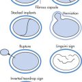
Aspergilloma (Mycetoma, Fungus Ball)
This is a saprophytic infection that occurs in the setting of structural lung disease (from TB, sarcoid, emphysema). Commonly in ULs, solitary lesions. The fungus grows into the preexisting cavity (e.g., cyst, bulla, bronchiectasis), creating a “fungus ball” consisting of fungus, mucus, and inflammatory cells. Individuals with mycetomas are often asymptomatic but may develop recurrent hemoptysis, which in rare cases can be massive. In these cases, bronchial artery embolization is indicated. The other treatment options include surgical resection, intracavity administration of amphotericin B, and systemic antifungal therapy.
Radiographic Features
- •
Focal intracavitary mass (3–6 cm), typically in ULs.
- •
Air may surround the aspergilloma (Monod sign), mimicking the appearance of cavitation seen with invasive aspergillosis.
- •
Small area of consolidation around cavity is typical.
- •
Adjacent pleural thickening is common.
- •
Fungus ball moves with changing position.
Invasive Aspergillosis
Invasive aspergillosis has a high mortality rate (70%–90%) and occurs mainly in severely immunocompromised patients (bone marrow transplants, leukemia). The infection starts with endobronchial fungal proliferation and then leads to vascular invasion with thrombosis and infarction of the lung (“angioinvasive infection”). Additional sites of infection (in 30%) are the brain, liver, kidney, and GI tract. Treatment is with systemic and/or intracavitary administration of amphotericin.
Radiographic Features ( Fig. 1.25 )
- •
Multiple pulmonary nodules, 40%
- •
Nodules have a characteristic halo of ground-glass appearance (represents pulmonary hemorrhage)
- •
Within 2 weeks, 50% of nodules undergo cavitation, which results in the air crescent sign. The appearance of the air crescent sign indicates the recovery phase (increased granulocytic response). The air crescent sign may also be seen in TB, actinomycosis, mucormycosis, septic emboli, and tumors. Do not confuse the air crescent sign with the Monod sign (clinical history helps to differentiate the two).
- •
Other manifestations:
Peribronchial opacities
Focal areas of consolidation

Semiinvasive Aspergillosis
This form of aspergillosis occurs in mildly immunocompromised patients and has a pathophysiology similar to that of invasive aspergillosis except that the disease progresses more chronically over months (mortality rate 30%). Risk factors include diabetes, alcoholism, pneumoconiosis, malnutrition, and COPD. Treatment is with systemic and/or intracavitary administration of amphotericin.
Radiographic Features
- •
Appearance similar to that of invasive aspergillosis
- •
Cavitation occurs at 6 months after infection
Cryptococcosis
Caused by Cryptococcus neoformans , which has a worldwide distribution and is ubiquitous in soil and pigeon excreta. Infection occurs through inhalation of contaminated dust.
Clinical Findings
Common in patients with lymphoma, diabetes, or AIDS and in patients receiving steroid therapy.
Radiographic Features
- •
Most common findings in the lung are pulmonary mass, multiple nodules, or segmental or lobar consolidation.
- •
Cavitation, adenopathy, and effusions are rare.
- •
Disseminated form: CNS, other organs
Candidiasis
Caused by Candida albicans more frequently than other Candida species.
Clinical Findings
Typically in patients with lymphoreticular malignancy; suspect pulmonary disease if associated with oral disease. Often there is disseminated fungemia.
Radiographic Features
- •
Plain radiograph is nonspecific: opacities (LL) > nodules.
- •
Nodular disease in disseminated form
- •
Pleural effusion, 25%
- •
Cavitation and adenopathy are rare.
Zygomycoses
Group of severe opportunistic mycoses caused by fungi of the Zygomycetes class:
- •
Mucormycosis ( Mucor )
- •
Rhizopus
- •
Absidia
Zygomycoses usually have two major clinical manifestations:
- •
Pulmonary mucormycosis
- •
Rhinocerebral mucormycosis
Zygomycoses are uncommon infections and occur primarily in immunocompromised patients (leukemia, AIDS, chronic steroid use, diabetes).
Radiographic Features
- •
Radiographic features are similar to those of invasive aspergillosis because of angioinvasive behavior of fungi.
Acquired Immunodeficiency Syndrome
General
AIDS is caused by HTLV type III (human T-cell lymphotrophic virus = HIV [human immunodeficiency virus]). HIV-1 and HIV-2 are single-stranded RNA viruses that bind to CD4 present on T lymphocytes (other cells: glial cells, lung monocytes, dendritic cells in lymph nodes). The viral RNA genome is copied into DNA with the help of reverse transcriptase and integrated into the host cellular DNA.
Known routes of HIV transmission:
- •
Blood and blood products
- •
Sexual activity
- •
In utero transmission
- •
During delivery
Clinical Findings
- •
Lymphadenopathy
- •
Opportunistic infections
- •
Tumors: lymphoma—usually B-cell non-Hodgkin’s lymphoma (NHL), Kaposi sarcoma (KS)
- •
Other manifestations:
Associated with lymphocytic interstitial pneumonia (LIP), usually in childhood
Spontaneous pneumothorax (development of cystic spaces, interstitial fibrosis related to PCP)
Septic emboli
- •
Clinical findings supportive of AIDS diagnosis ( Fig. 1.26 ):
CD4 cell count <200/mm 3 ; the dysfunction of the immune system is inversely related to the CD4 cell count; PCP, CD4 cell count <200/mm 3 ; MAI infection, CD4 cell count <50/mm 3
Less than one case of bacterial pneumonia per year

FIG. 1.26
Opportunistic Infections
- •
P. jiroveci Frenkel 1999, 70%
- •
Mycobacterial infection, 20%; CD4 cell counts often <50/mm 3
- •
Bacterial infection, 10% ( S. pneumoniae , Haemophilus )
- •
Fungal infection (<5% of AIDS patients)
- •
Nocardia, <5%: cavitating pneumonia
- •
CMV pneumonia (common at autopsy)
Chest
General
- •
50% of all AIDS patients have pulmonary manifestations of infection or tumor.
- •
A normal CXR does not exclude the diagnosis of PCP.
- •
CMV infection is common at autopsy but does not cause significant morbidity or death; CMV antibody titers are present in virtually all patients with AIDS.
- •
Use of chest CT in AIDS patients:
Symptomatic patient with normal CXR; however, patients will commonly first undergo induced sputum or bronchoscopy or be given empirical therapy for PCP.
To clarify confusing CXR findings
Workup of focal opacities, adenopathy, nodules
Spectrum of Chest Manifestations ( Fig. 1.27 )
Nodules
- •
KS (usually associated with skin lesions)
- •
Septic infarcts (rapid size increase)
- •
Fungal: Cryptococcus , Aspergillus
- •
Large opacity: consolidation, mass
- •
Hemorrhage
- •
NHL
- •
Pneumonia
- •
Linear or interstitial opacities
- •
PCP
- •
Atypical mycobacteria
- •
KS
- •
Lymphadenopathy
- •
Mycobacterial infections
- •
KS
- •
Lymphoma
- •
Reactive hyperplasia, rare in thorax
- •
Pleural effusion
- •
KS
- •
Mycobacterial, fungal infection
- •
Pyogenic empyema
- •

Pneumocystis Jiroveci Pneumonia (PJP) ( Figs. 1.28–1.30 )


General
P . jiroveci is an atypical fungus that can result in pneumonia in immunocompromised individuals. PJP affects individuals with AIDS, transplant recipients, and patients receiving long-term corticosteroid therapy. When PJP occurs in HIV-infected individuals, it is usually only after the CD4 count has decreased to less than 200/mm 3 . The diagnosis of PJP can be confirmed by bronchoalveolar lavage or presence of the organism in a sputum sample.
Radiographic Features
- •
Interstitial pattern, 80%
CXR: bilateral perihilar ground-glass opacities or thickened interstitium
HRCT: ground-glass opacities, interlobular septal thickening, and in some cases multifocal areas of consolidation
- •
Up to one-third of patients with PJP will have a normal CXR.
- •
Multiple cysts of various sizes are seen in one-third of patients with PJP and may cause spontaneous pneumothorax.
- •
UL P. jiroveci involvement is common, because aerosolized pentamidine may not get to ULs; UL disease may mimic TB, but the latter may have pleural effusions or lymphadenopathy, both of which are uncommon in PJP.
- •
Atypical patterns, 5%
Unilateral disease
Granulomatous inflammation resulting in focal nodules or masses
- •
PJP as a presenting manifestation of AIDS is decreasing in frequency because of trimethoprim-sulfamethoxazole prophylaxis.
Mycobacterial Infection
M. tuberculosis > M. avium-intracellulare (MAI) (this pathogen usually causes extrathoracic disease). CD4 cell count usually <50/mm 3 .
Radiographic Features
- •
Hilar and mediastinal adenopathy common
Necrotic lymph nodes (TB) have a low attenuation center and only the rim is enhanced with contrast medium.
M. tuberculosis is more commonly associated with necrosis from MAI.
Adenopathy in KS or lymphoma is enhanced uniformly.
- •
Pleural effusion
- •
Other findings are similar to those of non-AIDS TB (UL consolidations, cavitations).
Fungal Infections
Fungal infections in AIDS are uncommon (<5% of patients):
- •
Cryptococcosis (most common); 90% have CNS involvement.
- •
Histoplasmosis: nodular or miliary pattern most common; 35% have a normal CXR.
- •
Coccidioidomycosis: diffuse interstitial pattern, thin-walled cavities
Kaposi Sarcoma (KS) ( Fig. 1.31 )
The most common tumors in AIDS are:
- •
KS (15% of patients); incidence declining; male-female ratio 50 : 1
- •
Lymphoma (<5% of patients)

Pulmonary manifestations of KS (almost always preceded by cutaneous/visceral involvement):
- •
Nodules
1–3 cm
Spiculated “flame-shaped” nodules with peribronchovascular distribution in advanced disease
- •
Coarse reticular opacities emanating from hilum
- •
Pleural effusions (serosanguineous), 40%
- •
Adenopathy
- •
Lymphangitic tumor spread
AIDS-Related Lymphoma
NHL (usually aggressive B-cell type) > Hodgkin lymphoma.
Poor prognosis. The spectrum includes:
- •
Solitary or multiple pulmonary masses with or without air bronchogram, 25%
- •
AIDS-related lymphoma is typically an extranodal disease (CNS, GI tract, liver, bone marrow): adenopathy not very prominent.
- •
Pleural effusions are common.
Neoplasm
General
Location
Chest neoplasms are best categorized by their primary location:
- •
Lung tumors
- •
Pleural tumors
- •
Mediastinal tumors
- •
Tumors of the airway
- •
Chest wall tumors
Classification of Pulmonary Neoplasm
Malignant tumors
- •
Bronchogenic carcinoma
- •
Lymphoma
- •
Metastases
- •
Sarcomas, rare
- •
Low-grade malignancies (previously bronchial adenoma)
- •
Carcinoid, 90%
- •
Adenoid cystic carcinoma (previously cylindroma, resembles salivary gland tumor), 6%
- •
Mucoepidermoid carcinoma, 3%
- •
Pleomorphic carcinoma, 1%
- •
Benign tumors, rare
- •
Hamartoma
- •
Papilloma
- •
Leiomyoma
- •
Hemangioma
- •
Chemodectoma
- •
Pulmonary blastoma
- •
Chondroma
- •
Multiple pulmonary fibroleiomyomas
- •
Pseudolymphoma
- •
Lung Cancer Screening
- •
Low-dose chest CT
- •
Uses the Lung-RADS assessment to categorize patients, which can be accessed at https://www.acr.org/Quality-Safety/Resources/LungRADS .
- •
Lung-RADS stratifies patients on the basis of presence, size, and morphology of pulmonary nodules into different follow-up and management categories.
- •
Patients qualify if they are between 55 and 74 years old with at least a 30-pack-year smoking history.
- •
Yearly CT screening decreased lung cancer mortality in this subset of patients by 20% and all-cause mortality by 6.7% when compared with screening with radiography.
Percutaneous Biopsy
The true positive rate of percutaneous lung biopsy is 90%–95%. False-negative results are usually due to poor needle placement, necrotic tissue, and so on. Tumor seeding is extremely uncommon (1 in 20,000).
Contraindications to biopsy are usually relative and include:
- •
Severe COPD
- •
Pulmonary hypertension
- •
Coagulopathy
- •
Contralateral pneumonectomy
- •
Suspected echinococcal cysts
Technique
- 1.
Fluoroscopic or CT localization of nodule
- 2.
Pass needle over superior border of rib to avoid intercostal vessels
- 3.
Avoid traversing pulmonary veins and crossing fissure
- 4.
Coaxial needle system:
- •
19-gauge introducer needle
- •
20-gauge core biopsy needle with 1–2-cm needle throw
- •
- 5.
Cytopathologist should be present to determine if sample is adequate and diagnostic.
- 6.
CXR after procedure to determine presence of pneumothorax
Complications
- •
Pneumothorax, 25%; 5%–10% of patients require a chest tube (i.e., pneumothorax >25% or if patient is symptomatic)
- •
Hemoptysis
- •
Systemic air embolism: extremely rare but most serious complication, approximately 0.02%–0.7%
Bronchogenic Carcinoma
Bronchogenic carcinoma refers broadly to any carcinoma of the bronchus.
Classification
Adenocarcinoma (most common), 40%:
2011 International Association for the Study of Lung Cancer/American Thoracic Society/European Respiratory Society international multidisciplinary classification of lung adenocarcinoma:
- •
Adenocarcinoma in situ: ≤3 cm, noninvasive lepidic growth pattern (along the alveolar walls), typically nonsolid on CT, usually nonmucinous
- •
Minimally invasive adenocarcinoma: ≤3 cm, primarily lepidic growth with invasive component ≤5 mm, predominantly nonsolid appearance on CT but may contain central solid component (≤5 mm), typically nonmucinous
- •
Lepidic predominant nonmucinous adenocarcinoma: invasive but with mainly lepidic growth, typically part solid on CT but may have nonsolid or cystic appearance
- •
Invasive mucinous adenocarcinoma: predominantly lepidic growth pattern, formerly referred to as mucinous bronchioloalveolar carcinoma , may appear solid, primarily solid, part solid, or nonsolid on CT, can be multifocal
- •
Atypical adenomatous hyperplasia is one of the preinvasive lesions for adenocarcinoma of the lung:
- •
Typically a ground-glass nodule less than 5 mm in size
- •
SCC, 30%:
- •
Spindle cell carcinoma
- •
Small cell carcinoma, 15%:
- •
Oat cell
- •
Intermediate cell type
- •
Combined oat cell carcinoma
- •
Large cell carcinoma, 1%:
- •
Giant cell carcinoma
- •
Clear cell carcinoma
- •
Adenosquamous tumor
Risk Factors for Bronchogenic Carcinoma
- •
Smoking: 98% of male patients and 87% of female patients with lung cancer smoke; 10% of heavy smokers will develop lung cancer. The strongest relationship between smoking and cancer has been established for SCC, followed by adenocarcinoma.
- •
Radiation, uranium mining
- •
Asbestos exposure
- •
Genetic predisposition (HLA-Bw44 associated?)
Radiographic Spectrum
Primary Signs of Malignancy ( Fig. 1.32 )
- •
Mass (>6 cm) or nodule (<6 cm) with spiculated, irregular borders
- •
Unilateral enlargement of hilum: mediastinal widening, hilar prominence
- •
Cavitation
Most common in ULs or superior segments of LLs
Wall thickness is indicative of malignancy
Less than 4 mm: 95% of cavitated lesions are benign
>15 mm: 85% of cavitated lesions are malignant
Cavitation is most common in SCC
- •
Certain tumors may present as chronic ASD: adenocarcinoma, lymphoma
- •
Some air bronchograms are commonly seen by HRCT in adenocarcinoma.

Secondary Signs of Malignancy ( Fig. 1.33 )
- •
Atelectasis (Golden inverted S sign in RUL, LUL collapse)
- •
Obstructive pneumonia
- •
Pleural effusion
- •
Interstitial patterns: lymphangitic tumor spread
- •
Hilar and mediastinal adenopathy
- •
Metastases to ipsilateral, contralateral lung
| Tumor | Frequency (%) | Location | Comments |
|---|---|---|---|
| Adenocarcinoma | 40 | Peripheral | Scar carcinoma |
| Squamous cell carcinoma | 30 | Central, peripheral a | Cavitation |
| Small cell carcinoma | 15 | Central, peripheral a | Endocrine activity |
| Large cell carcinoma | 1 | Central, peripheral | Large mass |

Paraneoplastic Syndromes of Lung Cancer
Incidence: 2% of bronchogenic carcinoma
Metabolic
- •
Cushing syndrome (ACTH)
- •
Inappropriate antidiuresis (ADH)
- •
Carcinoid syndrome (serotonin, other vasoactive substances)
- •
Hypercalcemia (PTH, bone metastases)
- •
Hypoglycemia (insulin-like factor)
- •
Musculoskeletal
- •
Neuromyopathies
- •
Clubbing of fingers (HPO)
- •
Other
- •
Acanthosis nigricans
- •
Thrombophlebitis
- •
Anemia
- •
Radiation Pneumonitis
Radiation pneumonitis is the acute phase of radiation damage and usually appears 3 weeks after treatment. The minimum radiation dose to induce pneumonitis is 30 Gy. The acute phase is typically asymptomatic but may be associated with fever and cough. Fibrosis usually occurs after 6–12 months.
Radiographic Features
- •
Diffuse opacities in radiation port
- •
HRCT allows better assessment of extent than plain radiograph
Tumor Staging ( Fig. 1.34 )

American Joint Committee on Cancer Tumor-Node-Metastasis (TNM) Staging System, Seventh Edition (Nonsmall Cell Lung Cancer)
Primary tumor (T)
T0: No evidence of a primary tumor
T1: ≤3 cm, limited to lung, not more proximal than lobar bronchus
T1a: ≤2 cm
T1b: >2 and ≤3 cm
T2: >3 but ≤7 cm
- •
Or invades parietal pleura
- •
Or involves main bronchus ≥2 cm distal to carina
- •
Or atelectasis/obstructive pneumonia extending to hilum but not involving entire lung
- •
T2a: >3 but ≤5 cm
T2b: >5 but ≤7 cm
T3: >7 cm
- •
Or directly invades chest wall, diaphragm, phrenic nerve, mediastinal pleura, parietal pericardium
- •
Or mainstem bronchus <2 cm distal to carina
- •
Or atelectasis/obstructive pneumonia involving entire lung
- •
Or separate tumor nodules in a different ipsilateral lobe
- •
T4: Any size
- •
Heart, mediastinum, great vessels, trachea, recurrent laryngeal nerve, esophagus, vertebral body, or carina
- •
Or separate tumor nodules in a different ipsilateral lobe
- •
Nodes (N)
N0: No lymph node involvement
N1: Ipsilateral nodes in the hilar, interlobar, lobar, segmental, subsegmental regions
N2: Ipsilateral nodes in mediastinum. Affected regions include paratracheal, prevascular and retrotracheal, lower paratracheal, subcarinal, paraesophageal, and pulmonary ligament regions.
N3: Contralateral hilar or mediastinal nodes; all significantly large lymph nodes in the ipsilateral or contralateral supraclavicular or scalene regions
Metastases (M)
M0: No metastases
M1: Metastases
M1a: Intrathoracic metastases:
- •
Tumor nodules in contralateral lung
- •
Malignant pleural/pericardial effusion
- •
Pleural dissemination
- •
M1b: Distant (extrathoracic) metastases: liver, bone, brain, adrenal gland
Unresectable Stages
- •
Tumors are unresectable if T4, N3, or M1 (stage 3b or 4)
- •
Stage 3b: N3, M0, any T; T4, M0, any N ≥2
- •
Stage 4: M1, any T, any N
Summary of Treatment
- •
Tumors ≤5 cm without metastases can be treated surgically.
- •
Tumors ≤7 cm with an ipsilateral hilar metastasis and tumors either >7 cm or invading the pleura, chest wall, or diaphragm without metastases are treated with surgery followed by chemotherapy.
- •
Any tumor with ipsilateral mediastinal nodes, invasion of the mediastinum/mediastinal structures, and/or cancer in a different lobe is treated primarily with chemoradiation.
5-Year Survival
Overall 5-year survival rate is 15%.
Stage 1A: 50%
Stage 1B: 43%
Stage 2A (limited disease): 36%
Stage 2B (T4): 25%
Stage 3A (N3): 19%
Stage 3B: 7%
Stage 4: 2%
Small Cell Cancer Staging
Small cell lung cancer (SCLC) accounts for 15% of all lung cancers and is known for its rapid growth rate and its early dissemination to regional lymph nodes and distant sites.
Traditionally a two-stage system had been used for SCLC: “limited” and “extensive.” Limited disease is confined to one lung with ipsilateral lymph node metastasis (can be encompassed by a single radiation port) and has better prognosis. Extensive stage denotes metastases to the contralateral lung and nodes or distant organs (including the pleura). About two-thirds of patients with SCLC present at the extensive stage at the time of initial diagnosis. The International Committee for the Study of Lung Cancer now recommends that SCLC be staged with the eighth edition of the American Joint Committee on Cancer’s TNM staging system.
Five-year survival rates in patients with SCLC:
Stage 1A: 56%
Stage 1B: 57%
Stage 2A: 38%
Stage 2B: 40%
Stage 3A: 12%
Stage 3B: 0%
Lymph Node Imaging
Anatomy ( Fig. 1.35 )
Anterior mediastinal nodes
- •
Parietal node group
Internal mammary nodes
Superior diaphragmatic nodes
- •
Prevascular node group (anterior to the great vessels)
- •
Middle mediastinal nodes
- •
Paratracheal *
* Paratracheal and subcarinal nodes are best assessed by mediastinoscopy; the remainder of the nodes are best assessed by CT.
; the lowest node is the azygos node
- •
Subcarinal * : below bifurcation; drainage to right paratracheal nodes
- •
Subaortic; AP window node
- •
Tracheobronchial (pulmonary root, hilar)
- •
Posterior mediastinal nodes
- •
Paraaortic
- •
Prevertebral
- •
Paraspinous: lateral to vertebral body
- •

American Thoracic Society Classification ( Fig. 1.36 )
This classification system assigns numbers to regional lymph nodes:
- •
2R, 2L: paratracheal
- •
4R, 4L: superior tracheobronchial
- •
5, 6: anterior mediastinal
- •
7: subcarinal
- •
8, 9: posterior mediastinal
- •
10R, 10L: bronchopulmonary
- •
11R, 11L: pulmonary
- •
14: diaphragmatic

CT Criterion for Abnormal Nodes
Short-axis lymph node diameter >1 cm (60%–70% accuracy for differentiating between malignant and benign adenopathy)
Chest Wall Invasion
Accuracy for detection of chest wall invasion by CT is 40%–60%.
Radiographic Features ( Fig. 1.37 )
Reliable signs
- •
Soft tissue mass in chest wall
- •
Bone destruction
- •
Unreliable signs
- •
Obtuse angles at contact between tumor and pleura
- •
>3 cm of contact between tumor and pleura
- •
Pleural thickening
- •
Increased density of extrapleural fat
- •

Mediastinal Invasion
Contiguous invasion of mediastinal organs, heart, great vessels, aerodigestive tract, and vertebra indicates nonresectability.
Radiographic Features
- •
Diaphragmatic paralysis (phrenic nerve involvement)
- •
Mediastinal mass with encasement of mediastinal structures
- •
MRI may be useful to detect vascular invasion.
Malignant Pleural Effusion
Development of pleural effusions usually indicates a poor prognosis. Presence of a documented malignant pleural effusion makes a tumor unresectable (M1). Incidence of pleural effusion:
- •
Bronchogenic carcinoma, 50%
- •
Metastases, 50%
- •
Lymphoma, 15%
Pericardial Metastasis
Pericardial effusion with enhanced nodules is highly suggestive of malignant involvement of the pericardium.
CT and MRI findings are inconclusive for determination of benign versus malignant pleural and pericardial disease. Fluorodeoxyglucose positron emission tomography (FDG PET) has been shown to have high sensitivity in detecting pleural malignancy.
Pathogenesis of Malignant Effusions
- •
Pleural invasion increases capillary permeability.
- •
Lymphatic or venous obstruction decreases clearance of pleural fluid.
- •
Bronchial obstruction → atelectasis → decrease in intrapleural pressure.
Central Bronchial Involvement
Tumors that involve a central bronchus usually cause lung collapse or consolidation. These tumors are considered unresectable (T4 tumors) only if they involve the carina.
Metastases to Other Organs
Lung tumors most frequently metastasize to:
Liver (common)
Adrenal glands (common)
- •
30% of adrenal masses in patients with adenocarcinoma are adenomas.
- •
Most adrenal masses in patients with small cell carcinomas are metastases.
- •
Tumor may be present in a morphologically normal-appearing gland.
- •
Other sites (especially small and large cell tumors)
- •
Brain (common)
- •
Bones
- •
Kidney
- •
Specific Lung Tumors
Adenocarcinoma
- •
Now the most frequent primary lung cancer
- •
Typically presents as a multilobulated, peripheral mass
- •
May arise in scar tissue: scar carcinoma
- •
KRAS mutation is the most frequent genetic mutation.
Bronchioloalveolar Carcinoma
Subtype of adenocarcinoma; slow growth. The characteristic radiographic presentations are:
- •
Morphologic type
Small peripheral nodule (solitary form), 25% (most common)
Multiple nodules
Chronic ASD
- •
Air bronchogram
- •
Absent adenopathy
- •
Cavitation may be seen by HRCT (Cheerio sign)
- •
Squamous Cell Carcinoma (SCC)
SCC is most directly linked with smoking. SCC carries the most favorable prognosis. The most characteristic radiographic appearances are:
- •
Cavitating lung mass, 30%
- •
Peripheral nodule, 30%
- •
Central obstructing lesion causing lobar collapse
- •
Chest wall invasion
- •
FGFR1 amplification is the most common mutation.
Large Cell Carcinoma
Usually presents as large (>70% are >4 cm at initial diagnosis) peripheral mass lesions. Overall uncommon tumor.
Pancoast Tumor (Superior Sulcus Tumor)
Tumor located in the lung apex that has extended into the adjacent chest wall. Histologically, Pancoast tumors are often SCCs.
Clinical Findings
- •
Horner syndrome
- •
Pain radiating into arm (invasion of pleura, bone, brachial plexus, or subclavian vessels)
Radiographic Features ( Fig. 1.38 )
- •
Apical mass
- •
Chest wall invasion
- •
Involvement of subclavian vessels
- •
Brachial plexus involvement
- •
Bone involvement: rib, vertebral body

Small Cell Carcinoma (SCC) (Neuroendocrine Tumor, Type 3)
Most aggressive lung tumor with poorest prognosis. At diagnosis, two-thirds of patients already have extrathoracic spread:
- •
Typical initial presentation: massive bilateral lymphadenopathy
- •
With or without lobar collapse
- •
Brain metastases
Carcinoid (Neuroendocrine Tumor, Types 1 and 2)
Represent 90% of low-grade malignancy tumors of the lung. The 10-year survival rate with surgical treatment is 85%. Rarely results in carcinoid syndrome caused by the production of 5-hydroxytryptamine with flushing, diarrhea, nausea, and wheezing; carcinoid syndrome usually accompanied by liver metastases.
Types
- •
Typical carcinoid: local tumor (type 1)
- •
Atypical carcinoid (10%–20%): metastasizes to regional lymph nodes (type 2); liver metastases are very rare
Radiographic Features
PET negative
80% centrally located within main, lobar, segmental bronchi
- •
Segmental or lobar collapse (most common finding)
- •
Periodic exacerbation of atelectasis
- •
Endobronchial mass
- •
Very rarely located within the trachea (1%)
- •
Peripherally located carcinoid, 20%
- •
Pulmonary nodule
- •
May be enhanced with contrast medium
- •
| Findings | Typical Carcinoid | Atypical Carcinoid | LCNEC | SCLC |
|---|---|---|---|---|
| Demographic Features | ||||
| Mean patient age (years) | 40–49 | 50–59 | 60–69 | 70–79 |
| Association with smoking | No | Yes | Yes | Yes |
| Male-female ratio | 1 : 1 | 2 : 1 | >2.5 : 1 | >2.5 : 1 |
| Histopathologic Features | ||||
| Mitoses per 10 HPFs | <2 | 2–10 | >10 | >50 |
| Necrosis | No | Yes | Yes | Yes |
| Imaging Findings | ||||
| Central to peripheral ratio | 3 : 1 | 3 : 1 | 1 : 4 | 10–20 : 1 |
| Calcification or ossification | 30% | 30% | 9% | ≤23% |
| Extrathoracic metastases | 15% | 15% | 35% | 60%–70% |
| Enhancement | High; central or rim | High; central or rim | High | High with necrosis |
| FDG uptake on PET | Low | Low | High | High |
Hamartoma ( Fig. 1.39 )

Hamartomas are mesenchymal tumors and are the most common benign tumors of the lung composed of cartilage (predominantly), connective tissue, muscle, fat, and epithelial tissue. 85% are identified as solitary pulmonary nodules, 5%–15% are endobronchial. They typically occur in individuals older than 50 years and are more common in men.
Radiographic Features
- •
Well-circumscribed, often lobulated pulmonary nodule
- •
Calcification identified in only 10% of hamartomas <3 cm but seen in approximately 75% of hamartomas ≥5 cm in size
- •
“Popcorn” calcification classically associated with hamartomas, but may also show stippled pattern
- •
Slow growth: 0.5–5 mm/year
- •
HRCT can show intralesional fat (65%)
Carney Triad
Predominant in Young Women (<30 Years Old)
- •
Gastric smooth muscle tumors (epithelioid leiomyosarcoma)
- •
Functioning extra-adrenal paraganglioma
- •
Pulmonary chondromas
Tracheobronchial Papillomatosis
Radiographic Features
- •
Multiple, well-demarcated nodules that can grow
- •
Cavitate with 2–3-mm-thick walls
- •
Air fluid levels may develop
- •
Risk of SCC 15 years after diagnosis
Pulmonary Blastoma
- •
Predominant in males, poor prognosis
Radiographic Features
- •
Large peripheral mass that is well circumscribed
- •
May show pleural invasion and may metastasize
Lung Metastases From Other Primary Lesions
General
Pathways of metastatic spread from a primary extrathoracic site to lungs (in order of frequency):
- •
Spread via pulmonary arteries (PAs)
- •
Lymphatic spread (celiac nodes → posterior mediastinal nodes + paraesophageal nodes) and in lung parenchyma
- •
Direct extension
- •
Endobronchial spread
- •
Follow the invasion-metastasis cascade: invade locally, intravasate into blood, survive circulation, lodge in distant organ, merge into parenchyma, survive the foreign organ, and reinitiate growth.
- •
Neoplasms with rich vascular supply draining into systemic venous system:
- •
Renal cell carcinoma (RCC)
- •
Sarcomas
- •
Trophoblastic tumors
- •
Testis
- •
Thyroid
Neoplasms with lymphatic dissemination:
- •
Breast (usually unilateral)
- •
Stomach (usually bilateral)
- •
Pancreas
- •
Larynx
- •
Cervix
Other neoplasms with high propensity to localize in lung:
- •
Colon
- •
Melanoma
- •
Sarcoma
Radiographic Features
- •
Multiple lesions, 95%; solitary lesions, 5%
- •
Lung bases > apices (related to blood flow)
- •
Peripheral, 90%; central, 10%
- •
Metastases typically have sharp margins.
- •
Fuzzy margins can result from peritumoral hemorrhage (choriocarcinoma, chemotherapy).
- •
Cavitations are common in SCCs from head and neck primary lesions.
Calcified Metastases
Calcifications in lung metastases are observed in:
Bone tumor metastases
- •
Osteosarcoma
- •
Chondrosarcoma
- •
Mucinous tumors
- •
Ovarian
- •
Thyroid
- •
Pancreas
- •
Colon
- •
Stomach
- •
Metastases after chemotherapy
Giant Metastases (“Cannon Ball” Metastases) in Asymptomatic Patients
- •
Head and neck cancer
- •
Testicular and ovarian cancer
- •
Soft tissue cancer
- •
Breast cancer
- •
Renal cancer
- •
Colon cancer
Sterile Metastases
Sterile metastases refers to pulmonary metastases under treatment that contain no viable tumor. Nodules typically consist of necrotic and/or fibrous tissue.
Chronic Lung Disease
Idiopathic Diseases
| Diagnosis | Clinical Findings | HRCT Features | Differential Diagnosis |
|---|---|---|---|
| UIP/IPF | 40–70 years, male > female; >6 month dyspnea, cough, crackles, clubbing; poor response to steroids | Peripheral, basal, subpleural reticulation and honeycombing ± ground-glass opacity | Collagen vascular disease, asbestosis, CHP, scleroderma, drugs (bleomycin, methotrexate) |
| NSIP | 40–50 years, male = female; dyspnea, cough, fatigue, crackles; may respond to steroids | Bilateral, patchy, subpleural ground-glass opacity, ± reticulation | Collagen vascular disease, CHP, DIP |
| RB-ILD | 30–50 years, male > female; dyspnea, cough | Ground-glass, centrilobular nodules, ± centrilobular emphysema | Hypersensitivity pneumonitis |
| AIP/diffuse alveolar damage | Any age, male = female; acute-onset dyspnea, diffuse crackles and consolidation | Ground-glass consolidation, traction bronchiectasis and architectural distortion | ARDS, infection, edema, hemorrhage |
| COP | Mean 55 years, male = female; <3 month history of cough, dyspnea, fever; may respond to steroids | Subpleural and peribronchial consolidation ± nodules in lower zones; atoll sign (ring-shaped opacity) | Collagen vascular disease, infection, vasculitis, sarcoidosis, lymphoma, alveolar carcinoma |
| DIP | 30–54 years, male > female; insidious onset weeks to months of dyspnea, cough | Ground-glass opacity, lower zone, peripheral | Hypersensitivity pneumonitis, NSIP |
| LIP | Any age, female > male | Ground-glass opacity, ± poorly defined centrilobular nodules, thin-walled cysts and air trapping | DIP, NSIP, hypersensitivity pneumonitis |
Related posts:


Stay updated, free articles. Join our Telegram channel

Full access? Get Clinical Tree


