KEY FACTS
Terminology
- •
Malignancy arising from intrahepatic bile duct (IHBD) or extrahepatic bile duct epithelium
Imaging
- •
Best diagnostic clue: Intra- or extrahepatic bile duct mass with upstream bile duct dilatation
- •
Intrahepatic cholangiocarcinoma
- ○
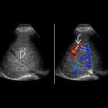
Mass with ill-defined margin, heterogeneous echotexture
- ○
Arise distal to 2nd-order bile ducts
- ○
Isolated thickening of IHBD or intraductal mass with upstream ductal dilatation
- ○
May see associated capsular retraction of liver
- ○
- •
Hilar cholangiocarcinoma
- ○
Dilatation of IHBDs without extrahepatic ductal dilatation
- ○
Nonunion of right and left hepatic ducts due to obstructing mass
- ○
Primary tumor may not be discernible or appears as small, infiltrative iso-/hyperechoic mass in hilar region
- ○
Nodular or polypoid mass in bile ducts
- ○
- •
Extrahepatic cholangiocarcinoma
- ○
Proportional bile duct dilatation
- –
Primary tumor often undetectable due to deep location
- –
- ○
Ill-defined, solid, heterogeneous mass within or surrounding duct at point of obstruction
- ○
Exophytic heterogeneous mass from bile duct
- ○
Polypoidal intraluminal tumor visible as iso-/hyperechoic mass within bile duct
- ○
Top Differential Diagnoses
- •
Pancreatic head adenocarcinoma
- •
Choledocholithiasis
- •
Recurrent pyogenic cholangitis
- •
Primary sclerosing cholangitis
- •
Porta hepatis tumor
Pathology
- •
Pathogenesis: Biliary intraepithelial neoplasm is considered premalignant lesion
Diagnostic Checklist
- •
Rule out other pancreatobiliary pathology causing biliary obstruction
- •
Long focal extrahepatic biliary stricture, irregular ductal wall, stenosis, and prestenotic biliary ductal dilatation
Scanning Tips
- •
Look for capsular retraction, biliary ductal dilatation, or abrupt cut-off of dilated bile ducts
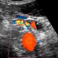
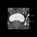
 at the confluence of the right and left hepatic ducts (Klatskin tumor). The mass invades adjacent liver parenchyma and hepatic veins
  , a common finding with cholangiocarcinoma. Note upstream dilatation of intrahepatic bile ducts
  .
.
Stay updated, free articles. Join our Telegram channel

Full access? Get Clinical Tree


