KEY FACTS
Terminology
- •
Choroid plexus cysts (CPC) are unilocular cysts usually located in the posterior thick portion of choroid plexus
Imaging
- •
CPC are most often incidental isolated findings
- ○
Almost all resolve by 32 weeks
- ○
Might be large, multiple, or bilateral
- ○
Not associated with aneuploidy in low-risk patients
- ○
- •
CPC may be seen in trisomy 18 (T18)
- ○
Anomalies and growth restriction almost always present
- –
Cardiac and extremity anomalies common
- –
- ○
CPC > 10 mm associated with mild ↑ risk for T18
- ○
Multiple and bilateral CPC do not ↑ risk for T18
- ○
Top Differential Diagnoses
- •
Intraventricular hemorrhage
- ○
Blood clot adherent to choroid might mimic CPC
- ○
Echogenic ventricular wall, ventriculomegaly
- ○
- •
Choroid plexus papilloma (rare)
- ○
Vascular mass, ventriculomegaly
- ○
Diagnostic Checklist
- •
Is patient low risk for aneuploidy? Has she had testing?
- ○
Cell-free DNA test results (best)
- ○
Maternal serum biochemistry results
- ○
Consider genetic counseling if patient has had no testing
- ○
- •
Follow-up to show CPC resolution is not necessary
Scanning Tips
- •
Do detailed scan of fetus when CPC is seen
- ○
Extra cardiac views
- –
Outflow and inflow tract views
- –
- ○
Extra extremity views
- –
Open hands, normal feet
- –
- ○
- •
Scan in multiple planes to show cyst located in choroid
- ○
Large cysts may be mistaken for ventriculomegaly
- ○
Normal fluid-filled atria may mimic CPC
- ○
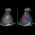
 occurring in the posterior, thick portion of the choroid plexus (glomus of choroid) is shown. The choroid plexus produces cerebrospinal fluid, and CPCs are considered “entrapment cysts”.


