Abstract
Monosomy for the distal portion of the short arm of chromosome 5 causes 5p deletion syndrome, which is also known by the currently less favored term “cri du chat” syndrome, from the French description of the monotonous high-pitched “cat-like” cry of affected infants. Initially described in 1963 by Lejeune et al., this syndrome is readily detectable by karyotype or with molecular cytogenetic methods, such as chromosomal microarray analysis (CMA). The prenatal diagnosis of this rare deletion syndrome is relatively uncommon. However, the recent introduction of noninvasive prenatal screening for microdeletion syndromes using cell-free fetal DNA (cffDNA) in maternal plasma has widened the prenatal ascertainment of 5p deletion syndrome to screening for potentially affected fetuses in low-risk pregnancies. Thus practitioners should be familiar with the clinical presentation and prognosis of 5p deletion syndrome, including features that can be seen by prenatal imaging and those that are not prenatally detectable, as well as with the implications for perinatal management and counseling when a 5p deletion is diagnosed prenatally.
Keywords
5p deletion, cri du chat, microcephaly, growth restriction, psychomotor delay
Introduction
Monosomy for the distal portion of the short arm of chromosome 5 causes 5p deletion syndrome, which is also known by the currently less favored term “cri du chat” syndrome, from the French description of the monotonous high-pitched “cat-like” cry of affected infants. Initially described in 1963 by Lejeune et al., this syndrome is readily detectable by karyotype or with molecular cytogenetic methods, such as chromosomal microarray analysis (CMA). The prenatal diagnosis of this rare deletion syndrome is relatively uncommon; however, recent introduction of noninvasive prenatal screening for microdeletion syndromes using (cffDNA) in maternal plasma has widened the prenatal ascertainment of 5p deletion syndrome to screening for potentially affected fetuses in low-risk pregnancies. Thus practitioners should be familiar with the clinical presentation and prognosis of 5p deletion syndrome, including features that can be seen by prenatal imaging and those that are not prenatally detectable, as well as with the implications for perinatal management and counseling when a 5p deletion is diagnosed prenatally.
Disorder
Definition
The 5p deletion syndrome is characterized by poor prenatal and postnatal growth, hypotonia, microcephaly, and a distinct round face with hypertelorism, micrognathia, epicanthal folds, and low-set ears. Affected children often have a high-pitched monotonous cry in infancy and have variable, but sometimes severe, psychomotor delay and intellectual disability.
Prevalence and Epidemiology
The 5p deletion syndrome is a rare genetic syndrome, affecting 0.2–0.6 : 10,000 individuals and is found in 0.3% to 1% of individuals with severe intellectual disability.
Etiology and Pathophysiology
The 5p deletion syndrome is caused by heterozygous partial deletions of the short arm of chromosome 5, which can be between 5 and 40 Mb. Most cases are the result of de novo terminal deletions (77%) or interstitial deletions (9%). For reasons incompletely understood, 80% of de novo deletions occur on the paternally inherited chromosome 5. The remainder are caused by unbalanced translocations, of which 5% are de novo and the remainder are familial, inherited from a parent carrying a balanced translocation (4%) or rarely an inversion (1%). Rare familial smaller deletions with significant variability in the phenotype within a family have been described, including one where a parent was ascertained after prenatal diagnosis of an affected fetus. Thus the degree of intellectual disability can be difficult to predict when an early diagnosis is made. A critical region from proximal 5p15.3 to distal 5p15.2 has been associated with the typical cry, and one in 5p15.2 is thought to be responsible for the dysmorphism, microcephaly, and intellectual disability. Genetic diagnosis can be achieved by standard G-banded karyotyping in many cases, and if the condition is clinically suspected, fluorescence in situ hybridization (FISH) with 5p15.2-specific probes can be performed. More recently, array-based copy-number analysis, or chromosomal microarray analysis (CMA) has become the method of choice for detection of microdeletions, including for prenatal diagnosis on amniotic fluid samples of pregnancies complicated by sonographically detected fetal anomalies. CMA has higher resolution and can achieve diagnosis for typical as well as for atypical presentations with smaller deletions and can differentiate simple deletions from unbalanced translocations or more complex genomic rearrangements. Noninvasive screening for microdeletion syndromes has recently become available but does not replace diagnostic testing in pregnancies complicated by fetal anomalies. Any positive results from such screening should be followed up by an offer for diagnostic testing by amniocentesis or CVS with CMA as the genetic testing strategy of choice. Finally, cases with mosaic deletions of 5p have been described, complicating genetic counseling.
Manifestations of Disease
Clinical Presentation
The typical clinical presentation of 5p deletion syndrome in infants includes low birth weight, microcephaly, a distinctive face, and the typical high-pitched cry, which is transient and related to delayed development of the larynx and hypotonia. Typical facial features include a round face with hypertelorism with a broad flat nasal bridge, epicanthal folds, down-slanting palpebral fissures, short philtrum, and micrognathia, most of which cannot easily be seen on prenatal imaging. Additional findings that occur less frequently include cardiac defects (present in up to 30% of cases), brain abnormalities (cerebellar hypoplasia, ventriculomegaly, and encephalocele), cystic hygroma, single umbilical artery, and hydrops. Several reports have shown that, although there is no specific predictive pattern, 5p deletion syndrome can be associated with high or low beta-human chorionic gonadotropin in several instances, as well as low maternal serum pregnancy associated plasma protein A (PAPP-a) or high alpha-fetoprotein on maternal serum analyte screening. Early in life it can be complicated by hypotonia, cyanotic crises, and feeding difficulties. Variable but often severe psychomotor delay becomes evident later. Affected individuals can survive into adulthood but have intellectual disability and often other neurobehavioral phenotypes (autistic features, and attention deficit hyperactivity disorder).
Imaging Technique and Findings
Ultrasound.
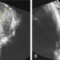
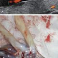
Table 156.1 lists the main prenatal sonographic findings of 5p deletion syndrome. The common US features of 5p deletion syndrome are fairly nonspecific and include microcephaly ( Fig. 156.1 ), growth restriction (both of which can have later onset after the second trimester), micrognathia ( Fig. 156.2 ), and hypertelorism ( Fig. 156.3 ). Some affected fetuses have been ascertained because of the presence of soft marker(s) for aneuploidy, including hypoplastic nasal bone ( Fig. 156.4 ), choroid plexus cysts ( Fig. 156.5 ), single umbilical artery, mild to moderate ventriculomegaly ( Fig. 156.6 ), thick nuchal fold, or cystic hygroma. Cardiac defects can be present in up to 30%. The most common are ventricular or atrial septal defects ( Fig. 156.7 ), patent ductus arteriosus (which cannot be diagnosed prenatally), followed by tetralogy of Fallot, pulmonary valve stenosis, or double-outlet right ventricle. While these cardiac defects are relatively rare in 5p deletion syndrome, they are overrepresented compared to the general population with congenital heart defects. Other rarer findings include encephalocele ( Fig. 156.8 ), facial clefting ( Fig. 156.9 ), cerebellar hypoplasia and Dandy-Walker malformation ( Fig. 156.10 ), hemivertebrae ( Fig. 156.11 ), and clinodactyly ( Fig. 156.12 ). All these findings are nonspecific, but when they occur, 5p deletion syndrome should be considered in the differential diagnosis. Conversely, if a 5p deletion is suspected by noninvasive cffDNA screening, ultrasound can be targeted to search for these features.
|


