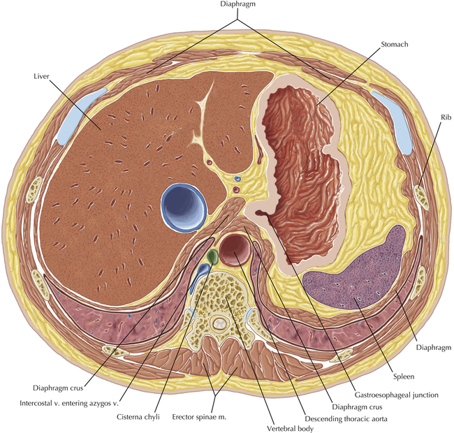
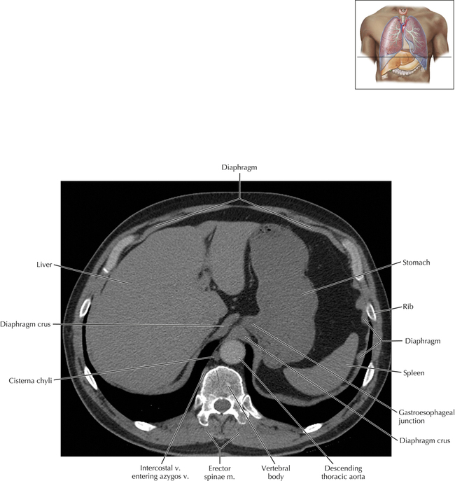
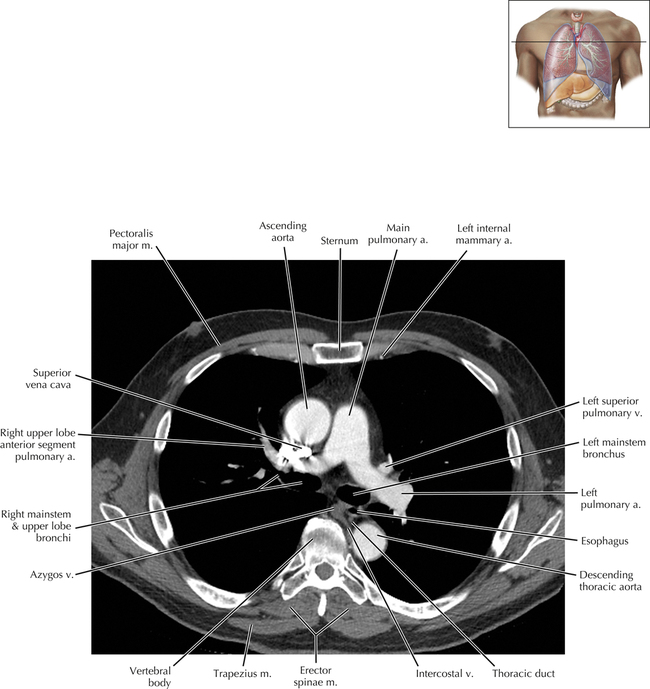
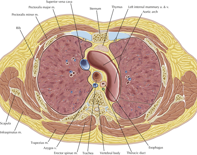
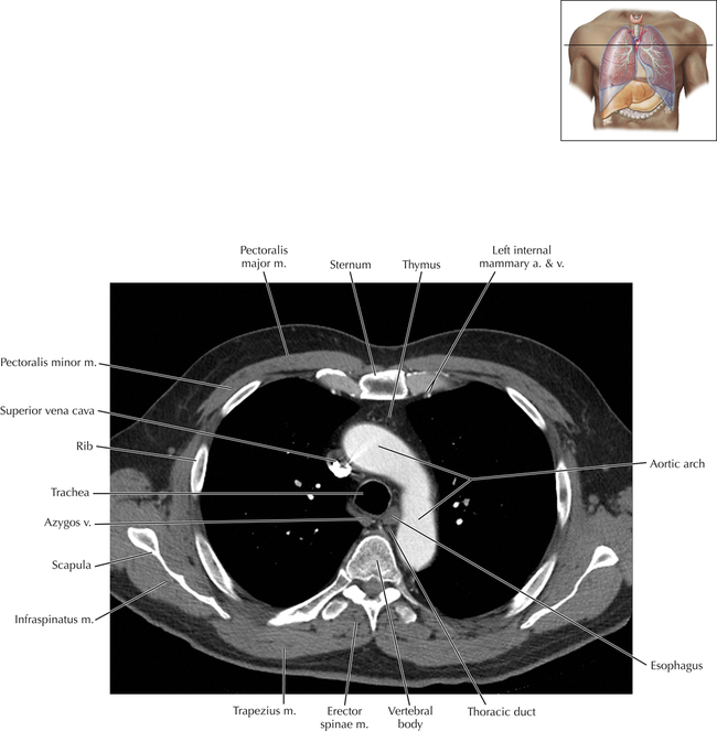
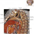
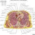
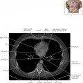
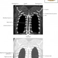
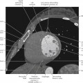
Chapter 5 Cisterna Chyli and Thoracic Duct AXIAL CORONAL Cisterna Chyli/Thoracic Duct Axial 1 Normal Anatomy A normal structure is shown—the cisterna chyli—which is inconsistently seen on clinical thoracic CT scans. The cisterna chyli is typically located between the distal thoracic or upper abdominal aorta and azygos vein in the right posterior mediastinum or retrocrural space, posterior to the right diaphragmatic crus. The cisterna chyli receives lymph from the right and left lumbar lymphatic trunks, the intestinal lymphatic trunk, and the inferior intercostal lymph nodes. The cisterna chyli gives rise to the thoracic duct, whose course will be shown on subsequent images. Cisterna Chyli/Thoracic Duct Axial 2 Normal Anatomy The thoracic duct is not always visible on thoracic CT scans, but is well seen in this patient. The thoracic duct originates from the cisterna chyli and ascends within the posterior mediastinum. The thoracic duct is well visualized adjacent to the azygos vein, posterior to the esophagus. Caudally, it usually ascends on the right side, often crossing to the left of midline at approximately the level of the 5th thoracic vertebral body. In this patient, the thoracic duct crossed midline at a more inferior level than is typically seen. Cisterna Chyli/Thoracic Duct Axial 3 Normal Anatomy The thoracic duct is again seen coursing within the posterior mediastinum adjacent to the azygos vein and descending thoracic aorta and posterior to the esophagus. Cisterna Chyli/Thoracic Duct Axial 4 Normal Anatomy At the level of the aortic arch at the cranial aspect of the azygos vein, the thoracic duct is again seen to the left of midline, posterior to the esophagus. Only gold members can continue reading. Log In or Register to continue Share this: Share on X (Opens in new window) X Share on Facebook (Opens in new window) Facebook Related posts: Overview of Cardiac Anatomy Venous Anatomy and Variants Overview of Thoracic Anatomy Thoracic Lymph Nodes Thoracic Soft Tissue and Lung Cardiac Anatomy Using CT Stay updated, free articles. Join our Telegram channel Join Tags: Netters Correlative Imaging Cardiothoracic Anatomy Dec 26, 2015 | Posted by admin in CARDIOVASCULAR IMAGING | Comments Off on Cisterna Chyli and Thoracic Duct Full access? Get Clinical Tree
Chapter 5 Cisterna Chyli and Thoracic Duct AXIAL CORONAL Cisterna Chyli/Thoracic Duct Axial 1 Normal Anatomy A normal structure is shown—the cisterna chyli—which is inconsistently seen on clinical thoracic CT scans. The cisterna chyli is typically located between the distal thoracic or upper abdominal aorta and azygos vein in the right posterior mediastinum or retrocrural space, posterior to the right diaphragmatic crus. The cisterna chyli receives lymph from the right and left lumbar lymphatic trunks, the intestinal lymphatic trunk, and the inferior intercostal lymph nodes. The cisterna chyli gives rise to the thoracic duct, whose course will be shown on subsequent images. Cisterna Chyli/Thoracic Duct Axial 2 Normal Anatomy The thoracic duct is not always visible on thoracic CT scans, but is well seen in this patient. The thoracic duct originates from the cisterna chyli and ascends within the posterior mediastinum. The thoracic duct is well visualized adjacent to the azygos vein, posterior to the esophagus. Caudally, it usually ascends on the right side, often crossing to the left of midline at approximately the level of the 5th thoracic vertebral body. In this patient, the thoracic duct crossed midline at a more inferior level than is typically seen. Cisterna Chyli/Thoracic Duct Axial 3 Normal Anatomy The thoracic duct is again seen coursing within the posterior mediastinum adjacent to the azygos vein and descending thoracic aorta and posterior to the esophagus. Cisterna Chyli/Thoracic Duct Axial 4 Normal Anatomy At the level of the aortic arch at the cranial aspect of the azygos vein, the thoracic duct is again seen to the left of midline, posterior to the esophagus. Only gold members can continue reading. Log In or Register to continue Share this: Share on X (Opens in new window) X Share on Facebook (Opens in new window) Facebook Related posts: Overview of Cardiac Anatomy Venous Anatomy and Variants Overview of Thoracic Anatomy Thoracic Lymph Nodes Thoracic Soft Tissue and Lung Cardiac Anatomy Using CT Stay updated, free articles. Join our Telegram channel Join