Fig. 27.1
Physical setup and components of three-dimensional (3D) visualization preoperative treatment planning system. Legend: ① a control unit of the electromagnetic tracking system ② two sensor interface devices of the electromagnetic tracking system ③ a field generator of the electromagnetic tracking system ④ tracked ultrasound probe ⑤ a simulation model ⑥ screen
27.3 Image Guidance Software and Tumor Segmentation
The image guidance software package was developed by our research team. The software provides the basic components needed for rapid prototyping and the development of image-guided microwave ablation applications. The graphical user interface displays the real-time simulation ultrasound 2D guided planning and the 3D visualization planning, as well as the planning path from the transverse, coronal, and sagittal plane. The tumor is segmented in a semiautomatic manner using the open source program FITME (http://www.fitme.org). The same user interface is also employed to allow the interventional radiologist to contour the tumor. Additionally, the system could visually display the tumor size and the distance between the tumor and the surrounding pipeline structure and organization.
27.4 Function of 3D Visualization Navigation System
Preoperative planning could be performed based on not only the 2D imaging but also 3D visualization model using this system, which could avoid the shortcomings of 2D imaging planning. Our approach to covering the entire tumor with a series of ablation spheres is based on the following guiding objectives: (1) covering the entire tumor plus a predefined ablative margin with a composite of spherical thermal fields, (2) minimizing the number of ablations, (3) minimizing the number of electrode insertion trajectories, and (4) avoiding any critical organ transgression or ablation. The planning software is designed to iterate through these goals until a complete treatment plan was finalized.
3D visualization navigation system combined with 3D visualization and navigation technology, focusing on the research of navigation parameters interface and image registration fusion. This system achieves the fusion of 2D ultrasound image and 3D visualization graphics and the display of navigation parameters interface, and it could be applied in image-guided intraoperative positioning in thermal ablation. In addition, this system could execute the preoperative planning accurately and improve the positioning capability of beginners by using it.
The quantitative assessment of postablation has always been a difficult point. 3D visualization navigation system could intuitively and quantitatively show the relationship between the tumor and ablation zone after ablation, realize quantitatively assessing ablation efficacy. The results of our simulated experiments showed that the accuracy rate of estimating tumor ablation efficacy by using this system was significantly higher than that by using conventional 2D imaging method.
27.5 Clinical Application of 3D Visualization Navigation System
3D visualization techniques can be used in every step of microwave ablation for liver cancer, including preoperative planning, intraoperative positioning, and postoperative assessment. We will introduce separately the clinical application of 3D visualization navigation system in three steps as follows.
27.5.1 Preoperative Planning
Preoperative treatment planning as the first step in the thermal ablation process acts to lower the rate of complications, ensure tumor-free safety margins after ablation, and improve long-term survival outcomes. The procedure of application of 3D visualization navigation system in preoperative planning is as follows: first, 3D visualization images are acquired according to the CT imaging data of patients in DICOM format; second, processing and analysis of the CT images is performed, and simulated ultrasound images are acquired; third, image registration is performed; fourth, doctors perform the preoperative planning using 3D visualization preoperative treatment planning system. The planning results are completed, including the number of planning insertions, the depth of insertions, angle of needles, and so on.
Liu et al. [15] reported the clinical application of 3D visualization preoperative planning in microwave ablation in liver cancer. Ninety-four enrolled patients with liver cancer were divided into two groups. The 3D preoperative planning group included 36 patients with 44 lesions, who underwent microwave ablation with the aid of the self-developed 3D visualization navigation system. The 2-dimensional (2D) preoperative planning group included 58 patients with 64 lesions, who underwent microwave ablation according to conventional 2D image preoperative planning method. After microwave ablation, therapeutic efficacy was assessed by contrast-enhanced imaging during follow-up. Results showed that the 3D preoperative planning group has a higher successful rate of first ablation than 2D preoperative planning group (p = 0.01). The sessions are more in 2D preoperative planning group than that in 3D preoperative planning group (p = 0.002). There were no significant differences in technique effectiveness rate between the 2D preoperative planning group (96.55 %) and 3D preoperative planning group (100 %) according to the contrast-enhanced images follow-up after microwave ablation (p = 0.64). There were no significant differences in the rate of local tumor progression between the 2D preoperative planning group and 3D preoperative planning group (p = 0.64) during 3–12 months’ follow-up (median, 6 months). Compared with 2D preoperative planning group, the 3D preoperative planning group has a higher successful rate of first ablation and less number of sessions. The application of 3D visualization preoperative planning system reduced the number of sessions compared with conventional 2D imaging preoperative planning method. Less session of ablation means less risk and less cost of hospitalization and shorter hospital stay, so the application of 3D visualization preoperative planning system is beneficial to patients with liver tumors. Meanwhile, based on the results, the 3D preoperative planning improved the accuracy of the positioning and may strengthen doctors’ confidence in ablation therapy. Therefore, the 3D visualization navigation system has a relatively high clinical application value in preoperative treatment planning of microwave ablation for liver cancer (Fig. 27.2).

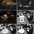
Fig. 27.2
Images in a 45-year-old patient with a tumor in the caudate lobe. (a) Preoperative CT image showed that the tumor (red arrows) was close to portal vein. (b) The 3D visualization images visualized the spatial relationship of tumor and the surrounding pipe in multi-angle. (yellow arrow: hepatic vein; green arrow: tumor; red arrow: portal vein) (c) The preoperative planning was achieved through 3D visualization of preoperative planning system, and three needles (white arrow) were needed to ablate the tumor completely. (d) The contrast-enhanced CT showed complete tumor necrosis (red arrows) a month after microwave ablation (MWA)
27.5.2 Intraoperative Positioning
Intraoperative positioning is the second key step of microwave ablation for liver cancer. Precise intraoperative positioning could be realized by using the navigation techniques. The 3D visualization navigation system achieved the fusion of 2D ultrasound image and 3D visualization graphics and the display of navigation parameters interface. This system could execute the preoperative planning accurately and improve the positioning capability of beginners by using it. The 3D visualization navigation system has the advantage of commercial navigation system; in addition, 3D visualization image will provide more information for microwave ablation and enhance ablation efficacy and decrease the complication.
Related posts:
 Microwave Ablation Combined with Cellular Immunotherapy for Hepatocellular Carcinoma
 Microwave Ablation for Malignant Liver Tumors Adjacent to the Hepatic Hilum
 Microwave Ablation of Large (≥5.0 cm) Hepatocellular Carcinoma
 Effectiveness of Contrast-Enhanced Ultrasound in Evaluating Microwave Ablation of Renal Cell Carcinoma
 Microwave Ablation in Other Tumors (Lung, Breast, and Bone)
 Microwave Ablation in Renal Cell Carcinoma
Stay updated, free articles. Join our Telegram channel

Full access? Get Clinical Tree


