In all, 1,574 simulated measurements of cervical diameter were obtained from 102 different examiners. Overall accuracy for determining the exact diameter was 56.3%, but improved to 89.5% when an error of ±1 cm was allowed. The intraobserver variability for a given measurement was 52.1% and decreased to 10.5% when an error of ±1 cm was allowed. The authors thus concluded that intraobserver variability is an important consideration in evaluating dysfunctional labor.
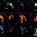
An electronic search in PubMed (1966 – December 2011), using the keywords “cervical dilatation,” “digital examination,” and “accuracy,” found only one article comparing the accuracy of ultrasound and vaginal examination for determining cervical dilatation during labor. In 2009, Zimerman et al. assessed the accuracy and reproducibility of intrapartum translabial three-dimensional (3D) ultrasonographic measurements of cervical dilatation during labor in a prospective observational study [2].
They collected 3D ultrasonographic volume data sets from 52 patients during labor and stored them. The correlation between the digital assessments by delivery room personnel and the digital vaginal examinations was studied, and interobserver and intraobserver agreement were determined. Translabial 3D ultrasonographic measurements of the mean and maximum cervical diameters and inner cervical area correlated positively with the digital findings (p < 0.001) with interobserver and intraobserver intraclass correlation coefficients of 0.82 and 0.85, respectively. The authors concluded that the assessment of cervical dilatation with 3D ultrasonography during labor is feasible and reproducible. However, this study did not demonstrate any benefit to the use of ultrasound. Therefore, despite the imperfect accuracy of digital examination for evaluating cervical dilatation, no tool is currently more accurate.
1.3 Evaluation of Fetal Head Station
Digital examination is also the criterion standard for the evaluation of fetal head station. Barbera et al. specifically addressed its accuracy for this purpose [3].
They developed a method for the objective assessment of fetal station and progression through the birth canal, based on simultaneous imaging by transperineal ultrasound (TPU) of both the maternal symphysis pubis and the fetal head.
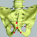
Since the true level of the ischial spines is critical to the assessment of clinical station, they developed a computed tomography (CT) geometric model to determine the angle between two vectors – the long axis of the pubic symphysis and a line drawn downward from the inferior margin of the symphysis – that best corresponds to the midpoint of a line drawn between the two ischial spines (representing clinical station 0). They showed that the mean angle (standard deviation) between these vectors was 99° (6°).
They next built an algorithm to associate each clinical station defined by the American College of Obstetricians and Gynecologists with a specific set of theoretical angles by creating consecutive nonoverlapping intervals around the theoretical mean (Table 1.1).
Table 1.1
Intervals of angle of head descent, derived from the geometric model and computed tomography data, associated with each specific fetal head station
Station | Lower angle (°) | Mean angle (°) | Upper angle (°) |
|---|---|---|---|
−5 | 62 | 65 | 68 |
−4 | 69 | 71 | 74 |
−3 | 75 | 78 | 81 |
−2 | 82 | 85 | 88 |
−1 | 89 | 92 | 95 |
0 | 96 | 99 | 102 |
1 | 103 | 106 | 109 |
2 | 110 | 113 | 116 |
3 | 117 | 120 | 123 |
4 | 124 | 127 | 131 |
5 | 132 | 135 | 139 |
For example, at station 0, the theoretical mean is 99°. Its lower angle was calculated as the midpoint between 92° (−1 station) and 99° (0 station), namely, 95°; its upper angle was calculated as the midpoint between 99° (0 station) and 106° (+1 station), namely, 102°. Finally, Barbera et al. assessed how closely clinical estimates of station, by digital examination, compared with ultrasound station by TPU in 88 laboring patients.
Figure 1.2 shows the relations between digitally assessed fetal head station and the TPU-recorded angles for each clinical station between −2 and +2 as well as their relation to the geometric model created with the CT data.
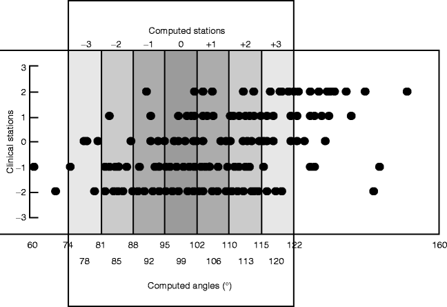

Fig. 1.2
Relationship between digitally assessed fetal head station and angle of head descent measured on transperineal ultrasound (•). Also shown are intervals of angle of head descent (shaded boxes) determined, using a geometric model, to be associated with each specific fetal head station (Barbera et al. [3])
Table 1.2 shows the extent of agreement between station determined by digital examination and that assigned by the geometric model. The highest percentage of complete agreement was only 46%, at −2 station. The percentage of complete agreement declined progressively from their downward. At computed station 0, for example, the digital examination completely agreed in only 18% of cases and for computed station +2, only 2.6%. On the other hand, station +2 as determined by digital examination was within ±2 cm of the computed station in 39% of cases.
Table 1.2
Agreement between the assessment of fetal head station by digital examination and by measurement of the angle of head descent, assessed by transperineal ultrasound
Agreement (%) | |||
|---|---|---|---|
Computed station | Complete | ±1 cm | ±2 cm |
−3 | 27 | 60 | 87 |
−2 | 46 | 92 | 100 |
−1 | 14 | 64 | 89 |
0 | 18 | 53 | 92 |
1 | 16 | 32 | 56 |
2 | 2.6 | 26 | 39 |
3 | 0 | 12 | 40 |
This result is certainly not surprising since the assessing fingers are able to feel both the spines and the fetal skull when the head is above the level of the spines. In contrast, once the fetal head is below 0 station, the ability to appreciate the relation between the ischial spines, located laterally in the pelvis, and the most prominent part of the centrally located fetal skull, presents a major challenge, demonstrated by progressively worsening agreement below 0 station. An agreement between 89% and 100% was observed only with a ±2-cm variation, that is, every time a clinician diagnoses the fetal head to be at 0 station, the real station may vary between −2 and +2. This inaccuracy is especially vexing at station +2.
As ACOG states that forceps application (low) is safe only at or after this station [4], it is rather critical.
Thus, each angle measured by TPU corresponded to a wide range of clinically assessed stations, and clinical digital assessment of station correlated poorly with computed station, especially at stations below zero, where the clinical impact could be substantial.
We must therefore search for more accurate tools to evaluate fetal head descent and to predict fetal head engagement. Digital examination should not be the criterion standard for evaluating these new tools.
1.4 Evaluation of Fetal Head Position
The data about intrapartum assessment of fetal head position are based on the time-honored X-ray study by Caldwell and Moloy [5] and a subsequent report by Calkins [6], both dating back to the1930s. Recent use of intrapartum ultrasound gives us the opportunity to address the accuracy of digital transvaginal examination in assessing fetal head position. Sherer et al. conducted two prospective studies to compare transvaginal digital examination and transabdominal ultrasound assessment [7, 8].
The first study was performed during the active stage of labor in 102 consecutive patients at term with normal singleton fetuses in cephalic presentation [7].
All participants had ruptured membranes; cervical dilation ≥4 cm and fetal head at ischial spine station −2 or lower. Transvaginal digital examinations were performed by either senior residents or attending physicians and followed immediately by transverse suprapubic transabdominal ultrasound assessments, considered to be the criterion standard. Examiners were blinded to each other’s findings. Transvaginal digital examinations were consistent with ultrasound assessments for only 24% of the patients (p = 0.002). Logistic regression revealed that cervical effacement (p = 0.03) and ischial spine station (p


Stay updated, free articles. Join our Telegram channel

Full access? Get Clinical Tree


