Etiology
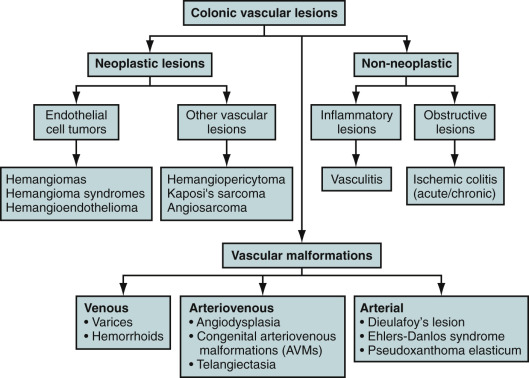
Vascular lesions of the colon are an important medical problem and have now been recognized as a significant cause of gastrointestinal bleeding. They can be solitary or multifocal, benign or malignant, or associated with a syndrome or systemic disorder. There are three main groups: vascular malformations, neoplastic lesions, and non-neoplastic lesions ( Figure 32-1 ). Vascular malformations can be broadly classified into arterial, venous, and arteriovenous types. Neoplastic lesions include hemangiomas, hemangioendotheliomas, and angiosarcomas. Non-neoplastic lesions can be further divided into inflammatory lesions (e.g., vasculitis) and obstructive lesions (e.g., ischemic colitis). Few systemic conditions and syndromes manifest with vascular lesions, such as colonic varices in portal hypertension or vasculitis in systemic lupus erythematosus, polyarteritis nodosa, Ehlers-Danlos syndrome, Osler-Weber-Rendu disease, Marfan syndrome, and systemic sclerosis. This chapter focuses on vascular lesions that cause gastrointestinal bleeding and that are representative of the spectrum of vascular lesions of the gastrointestinal tract.
Prevalence and Epidemiology
The prevalence is related to the cause and age of the patient. The prevalence of angiodysplasia is 0.8% in healthy patients older than 50 years who are undergoing screening colonoscopy. These lesions characteristically appear in the right colon and cecum in older patients, although they may be found anywhere in the lower gastrointestinal tract, may be multiple, and can occur in younger patients. Dieulafoy’s lesion involving the colon is rare and more frequently affects the stomach. It is twice as common in men as in women and manifests at a mean age of 52 years. Hemangiomas are benign vascular tumors that can be found throughout the gastrointestinal tract, often in the rectum or colon. The incidence of gastrointestinal hemangioma is reported as 0.3%, and these tumors account for 5% to 10% of all benign intestinal tumors. In some populations these lesions are multiple and associated with skin lesions, such as the blue rubber bleb nevus syndrome with purple-blue cutaneous hemangiomas or the Klippel-Trenaunay syndrome with port-wine–colored cutaneous hemangiomas, hemihypertrophy, and varicose veins. Rare vascular malignant neoplasms of the gastrointestinal tract include angiosarcomas, hemangiopericytomas, and hemangioendotheliomas. The incidence of these lesions is highly variable. Telangiectases are similar to angiodysplasias but occur in all the layers of the bowel wall, are usually congenital, and often occur in other organ systems. Hereditary hemorrhagic telangiectasia (Osler-Weber-Rendu disease) is an autosomal dominant disorder with telangiectases involving the lips; mucous membranes, especially in the mouth and nose; gastrointestinal tract, especially the stomach and small bowel; liver; lung; retina; and central nervous system.
Clinical Presentation
Although many vascular lesions are asymptomatic, those that manifest with bleeding require expeditious localization and control. These lesions may be responsible for severe, acute, and overt bleeding or alternatively can cause chronic or occult bleeding. Some of these lesions are difficult to localize on cross-sectional imaging and colonoscopy and necessitate use of more invasive modalities such as angiography. Although the clinical presentation, age of the patient, and other epidemiologic factors may indicate a particular type of colonic vascular lesion, endoscopy with biopsy and visceral angiography are critical in establishing the correct diagnosis.
Pathophysiology and Pathology
Pathologic processes of vascular lesions involving the colon depends on the underlying cause, which can be vascular, neoplastic, or non-neoplastic in origin.
Angiodysplasia
These lesions are acquired vascular ectasias, possibly caused by chronic, low-grade colonic obstruction. Histologic identification is difficult unless special techniques are used. Microscopically, angiodysplasias are composed of clusters of dilated, tortuous, thin-walled veins, venules, and capillaries localized in the colonic mucosa and submucosa.
Dieulafoy’s Lesion
Dieulafoy’s lesion belongs to an arterial type of vascular abnormality with abnormally large (caliber-persistent) submucosal end-arteries; in some instances, there is a small, overlying mucosal defect.
Congenital Arteriovenous Malformations
Histologically, arteriovenous malformations are persistent congenital communication between arteries and veins located in the submucosa. Characteristically, arterialization of the veins is seen. The veins are tortuous and dilated, having thick walls with smooth muscle hypertrophy and intimal thickening.
Hemangioma
Hemangioma is the second most common vascular lesion of colon and may be solitary or may be multiple in patients with multisystem involvement. Histologically, hemangiomas can be divided into cavernous, capillary, and mixed. Cavernous hemangiomas are composed of large, dilated spaces filled with blood and are covered with a thin wall of abnormal vessels. Capillary hemangiomas consist of conglomerates of small, thin-walled vessels.
Imaging
Because the clinical presentation and manifestations of patients with colonic vascular lesions in the form of lower gastrointestinal bleeding overlap with those of other colonic diseases, a precise identification of cause is possible only with a high degree of suspicion and by using different imaging modalities. A precise knowledge of clinical background with imaging findings can help narrow the differential diagnosis. Although there is no specific algorithm, the information presented in Box 32-1 and Table 32-1 should be kept in mind when there is a high index of suspicion of a vascular lesion.
- •
Age
- •
Elderly
- •
Angiodysplasia
- •
Dieulafoy’s lesion
- •
Ischemic colitis
- •
- •
Children
- •
Hemangiomas
- •
Hereditary hemorrhagic telangiectasia
- •
- •
- •
Location of Involvement
- •
Angiodysplasia: Cecum and proximal ascending colon
- •
Hemangiomas: Predilection for rectosigmoid region
- •
- •
Multifocal Involvement
- •
Hemangioma
- •
| Angiodysplasia | Hemangioma | Dieulafoy’s Lesion | |
|---|---|---|---|
| Age | Older than 50 years | Younger age group | No specific age distribution |
| Diagnostic modalities | Endoscopy, catheter angiography, or CT angiography | CT and endoscopy | Endoscopy and angiography |
| Radiographs | N/A | Phleboliths can be seen on radiographs. | N/A |
| CT/CTA Findings | Ectatic, dilated vessels in the wall of colon, an early filling vein, and an enlarged ileocolic artery | Enhancing lesion with phleboliths | N/A |
| MRI findings | N/A | Thickened colon wall with high signal intensity on T2-weighted imaging | N/A |
| Colonoscopy | Lesions are flat, 2-5 mm in diameter, and red. | Elevated blue nodular lesions or dilated vessels | Caliber-persistent artery protruding from the mucosa |
Angiodysplasia
Angiodysplasia or vascular ectasia is the most common vascular malformation of the gastrointestinal tract in the elderly and one of the major causes of lower gastrointestinal bleeding. Accurate diagnosis may require a combination of diagnostic techniques, such as angiography, nuclear scanning, and colonoscopy.
Barium Studies
Because the lesions of angiodysplasia are small in diameter and do not distort the mucosa, double-contrast barium enema studies are of no value in the diagnosis of angiodysplasia. This technique is useful, however, to rule out other causes of gastrointestinal bleeding, such as neoplastic lesions.
Computed Tomography
The role of CT is evolving, and at present there is limited literature on its role in evaluating angiodysplasia. There are some reports of being able to detect these lesions by multidetector CT (MDCT) scans and CT angiography (CTA) techniques. In a study of 30 patients with clinical suspicion of angiodysplasia, CTA had a sensitivity of 78% and a specificity of 100% when compared with colonoscopy, which had a sensitivity of 68% to 80% and a specificity of 90%. Accumulation of ectatic, dilated vessels in the colon wall, an early filling vein, and an enlarged ileocolic artery ( Figure 32-2 ) can be observed in CTA images.
Magnetic Resonance Imaging
The role of magnetic resonance imaging (MRI) is still investigational and evolving.
Nuclear Medicine
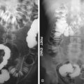
Nuclear scintigraphy is a sensitive method of detecting gastrointestinal bleeding at a rate of 0.1 mL/min. It is more sensitive than angiography but less specific than a positive endoscopic or angiographic examination ( Figure 32-3 ). A major disadvantage of nuclear imaging is that it localizes bleeding only to an area of the abdomen. Nuclear scintigraphy has proved more useful as an adjunct to angiography by localizing and confirming the presence of bleeding, minimizing the number of angiograms that do not yield meaningful diagnostic information, and allowing rapid selection of the artery to be injected by angiography.
Related posts:


Stay updated, free articles. Join our Telegram channel

Full access? Get Clinical Tree


