, Joon Woo Lee1 and Eugene Lee2
(1)
Department of Radiology, Seoul National University College of Medicine, Seoul National University Bundang Hospital, Seongnam, South Korea
(2)
Department of Radiology, Seoul National University Bundang Hospital, Seongnam, South Korea
The first step in the compartmental approach to spinal tumors is to determine if the lesion is intraosseous, extradural, intradural extramedullary (IDEM), or intramedullary (IM) in location. The possible differential diagnoses and optimal surgical approaches of these tumors are different depending on their compartmental location. In most cases it is not difficult to identify the compartment in which these tumors are situated on MRI, although it can be confusing in some instances. In the chapter, we will discuss radiological clues that can be helpful in localizing the compartment to which these tumors belong.
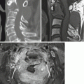
1.1 Extradural Versus Intradural Tumor
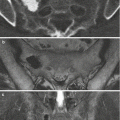
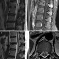
The main clues favoring an extradural tumor are extrinsic dural sac compression and tumor extension into the neural foramen. With severe tumor compression of the dural sac, however, it can be difficult to tell if the dural/cord compression is extrinsic (by an extradural tumor) or due to a space occupying lesion within the dura (by an intradural tumor). The clue that points toward an extradural tumor lies in the appearance of the subarachnoid space at the margin of the tumor; due to extrinsic compression of the dura, the subarachnoid space between the dura and spinal cord is obliterated at its margin of the tumor. With an intradural tumor, the dura bulges out instead with a convex appearance, while the cord is compressed by the tumor, resulting in widening of the subarachnoid space at the edge of the tumor.
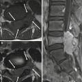
1.2 Intradural Extramedullary Versus Intramedullary Tumor
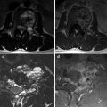
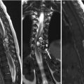
The clue that can be used to differentiate an extramedullary from an intramedullary tumor lies in the appearance of the outer contour of the spinal cord at the margin of the tumor. With an extramedullary tumor, the outer contour of the spinal cord at the interface with the tumor is indented upon and compressed by the tumor resulting in widening of the subarachnoid space, whereas with an intramedullary tumor the outer contour of the spinal cord has a convex bulge resulting in narrowing of the subarachnoid space.
Exophytic intramedullary tumors can occasionally be confusing and mimic appearances of extramedullary tumors, especially in the lower thoracic spinal cord and conus medullaris. In such cases, the axial images have to be reviewed carefully. With intramedullary tumors part of the tumor can be shown to be contiguous with the spinal cord, and if this can be established, it can be concluded that the tumor originates from the spinal cord.
Related posts:
Stay updated, free articles. Join our Telegram channel

Full access? Get Clinical Tree


