Diagnostic Ophthalmic Ultrasound for Radiologists 327
Cynthia J. Kendall, Thomas C. Prager, Han Cheng, Dan Gombos, Rosa A. Tang, and Jade S. Schiffman
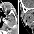
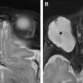
Ophthalmic ultrasound is an invaluable tool that provides quick and noninvasive evaluation of the eye and the orbit. It not only allows the clinicians to view structures that may not be visible with routine ophthalmic equipment or neuroimaging techniques but also provides unique diagnostic information in various ophthalmic conditions. In this article, the basic principles of ophthalmic ultrasound and examination techniques are discussed. Its clinical application is illustrated through a variety of ocular pathologic abnormalities (eg, narrow angles, ciliary body tumor, detached retina, choroidal melanoma, and papilledema).
Optical Coherence Tomography for the Radiologist 367
Jade S. Schiffman, Nimesh B. Patel, Roberto Alejandro Cruz, and Rosa A. Tang
Optical coherence tomography is an imaging technique using low coherence light sources to produce high-resolution cross-sectional images. This article reviews pertinent anatomy and various pathologies causing optic atrophy (eg, compressive, infiltrating, demyelinating) versus optic nerve swelling (from increased intracranial pressure known as papilledema or other optic nerve intrinsic pathologies). On optical coherence tomography, optic atrophy is often associated with reduced average retinal nerve fiber layer thickness, whereas optic nerve swelling is usually associated with increased average retinal nerve fiber layer thickness.
Advanced MR Imaging of the Visual Pathway 383
Fang Yu, Timothy Duong, and Bundhit Tantiwongkosi
Vision is one of our most vital senses, deriving from the eyes as well as structures deep within the intracranial compartment. MR imaging, through its wide selection of sequences, offers an array of structural and functional imaging tools to interrogate this intricate system. This review describes several advanced MR imaging sequences and explores their potential clinical applications as well as areas for further development.
Related posts:


Stay updated, free articles. Join our Telegram channel

Full access? Get Clinical Tree


